

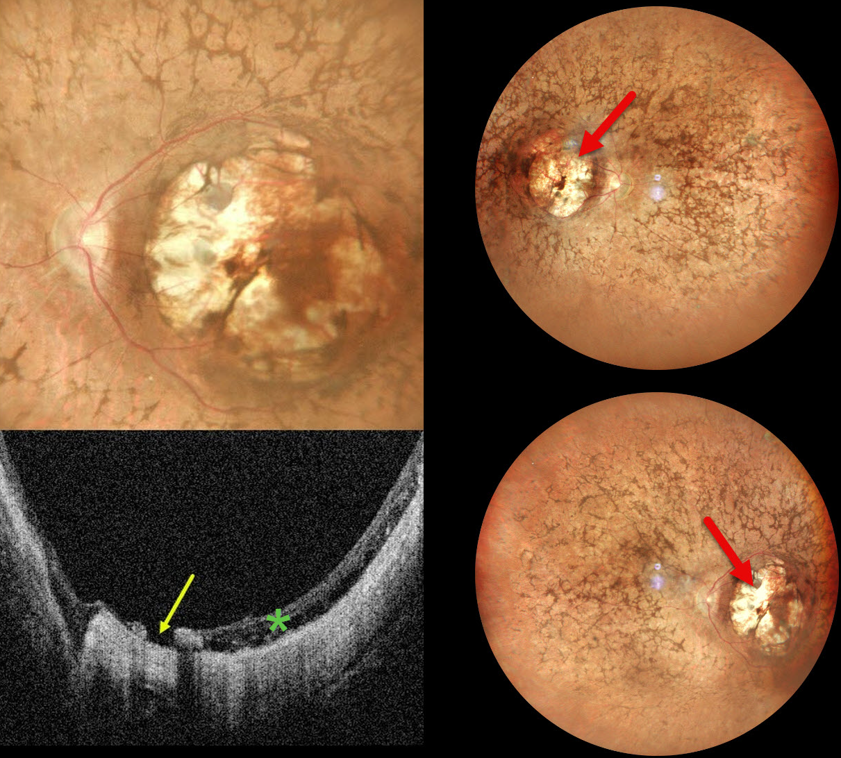
This 32YO female was born with severe vision loss and nystagmus. There was no family history of eye disease. Vision was counting fingers bilaterally.
Pseudocolor SLO imaging shows bilateral symmetric macular colobomas with diffuse retinitis pigmentosa (RP) findings, including vascular narrowing, diffuse pigmentary changes, and intraretinal pigment migration (bone spicules). OCT shows thinned and variably disorganized retinal bands within the colobomas, and a small macular hole is noted nasally OS.
Learning Points:
Macular colobomas are rarely associated with RP (Parmeggiani et al, Eye 2004;18:421-428). North Carolina macular dystrophy, caused by a mutation in the PRDM13 gene (Small et al, Ophthalmology 2016;123:9-18), is characterized by macular coloboma-like lesions. However, these eyes do not have peripheral RP findings. Genetic testing in our patient was not performed, and observation was recommended.



