

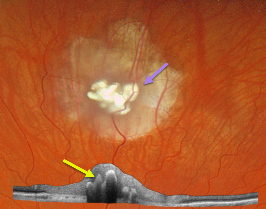
This healthy 30YO male was referred for an asymptomatic retinal lesion in his left eye. There was no family history of eye disease. Vision was 20/25 OU.
Optos color RGB imaging shows a nodular, calcified retinal lesion with surrounding atrophic chorioretinal scarring. Topcon swept-source OCT scanning shows a disorganized retina overlying the hyperreflective calcifications, which shadow posteriorly.
Learning Points:
Retinocytomas are fleshy, opalescent, benign tumors that are felt to represent spontaneously regressed retinoblastoma (RB). They appear identical to previously radiated RB tumors. As with typical RB, these patients’ offspring are at 50% risk of developing RB and thus require genetic counseling.
Clinically, retinocytomas can resemble astrocytic hamartomas. However, retinocytomas often have underlying RPE changes and chorioretinal scarring (as in our patient), which are not present in astrocytic hamartomas.
Brain MRI scanning was normal. Genetic testing for the retinoblastoma (RB1) and tuberous sclerosis (TSC1/TSC2) genes was suggested, but the patient denied this for now due to cost concerns.



