

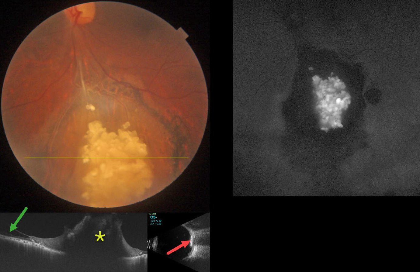
This healthy 43YO male presented with gradual bilateral blurring. Vision was 20/20 in his normal OD and 20/30 OS.
Color photography shows an elevated, glistening, inner retinal yellow-white mulberry-like lesion in the inferior midperiphery. Surrounding subretinal pigmentary changes are noted. OCT through this lesion shows a markedly thickened, disorganized, hyperreflective retina. Retinoschisis is noted nasally. The lesion is markedly hyperreflective on B-scan ultrasonography and hyper-autofluorescent on fundus autofluorescence.
Learning Points:
Retinocytomas are fleshy, opalescent, benign tumors that are felt to represent spontaneously regressed retinoblastoma (RB). They appear identical to previously radiated RB tumors. The risk of a germline RB1 mutation in sporadic unilateral retinoblastoma or retinocytoma is about 10-20%.
Clinically, retinocytomas can resemble astrocytic hamartomas. However, retinocytomas often have underlying RPE changes and chorioretinal scarring (as in our patient), which would not be present with astrocytic hamartomas.
The brain MRI was normal, and observation was recommended.



