

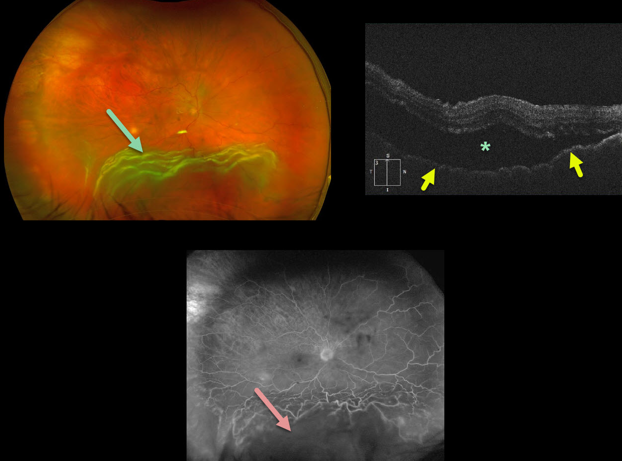
This 62YOM presented with vision loss from waxing and waning shifting unilateral subretinal fluid with marked chorioretinal folds and thickened choroid on OCT.
We initially agreed with the likely diagnosis of posterior scleritis that was suggested by a retinal specialist out of state.
However, fluorescein angiography showed no leakage and he failed to improve with a trial of oral prednisone.
Subsequent vitrectomy found a tiny tear superonasally and cytology was negative for lymphoma. His retina has remained attached since.
Learning Points:
The normal neurosensory retina remains attached to the RPE due to numerous physiologic mechanisms including the the inner (retinal vascular endothelium) and outer (tight junctions at the apex of the RPE cells) blood retinal barriers, hyperosmotic choroid passively drawing fluid into the subretinal space, RPE pump, interphotoreceptor matrix, and friction between the RPE microvilli and the photoreceptor outer segments.
Rhegmatogenous retinal detachment (RRD) occurs when liquid vitreous, entering the subretinal space through a retinal break, overwhelms these forces favoring retinal detachment.
Although shifting subretinal fluid is usually a sign of exudative detachment, it may also occur in RRD.



