

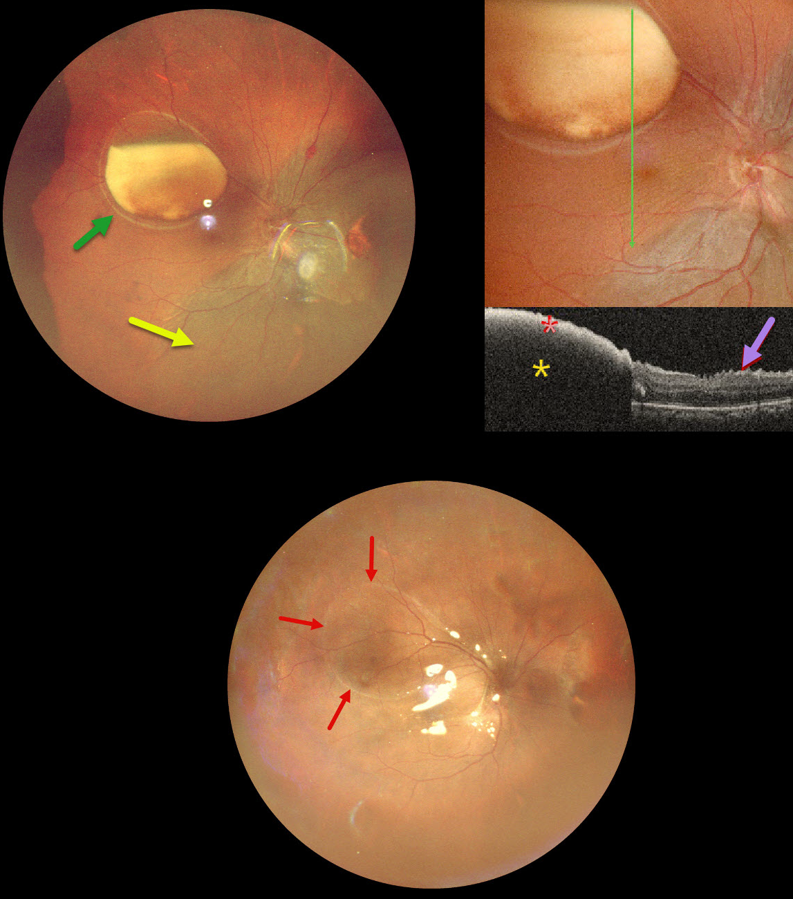
This 39YO male underwent vitrectomy OD elsewhere for a vitreous hemorrhage one month earlier. The treating surgeon noticed a sub-internal limiting membrane (ILM) hemorrhage at a postoperative visit and referred him to us for further evaluation. Vision was 20/30 OD and 20/20 in his normal OS.
Ultrawidefield pseudocolor SLO imaging shows a large partially layered sub-ILM hemorrhage with devitalized yellow blood in the superotemporal midperiphery. A retinal detachment is noted inferiorly and nasally. Inner retinal folds are visible at higher magnification. The blood is highly reflective on OCT scanning with total posterior shadowing. The inner retinal folds are noted inferiorly.
We don’t have a good explanation for our patient’s sub-ILM hemorrhage following vitrectomy for a vitreous hemorrhage. The yellow devitalized blood must have been present preoperatively, even though the initial surgeon told us he just noticed it one month postoperatively. Possibly our patient developed a breakthrough vitreous hemorrhage from a Valsalva maneuver, but without further history, this is just speculative.
We repaired the detachment with vitrectomy and silicone oil, and drained the sub-ILM blood intraoperatively. The retina was completely attached 2 weeks later. A high-water mark defines the margins of the prior ILM detachment.



