

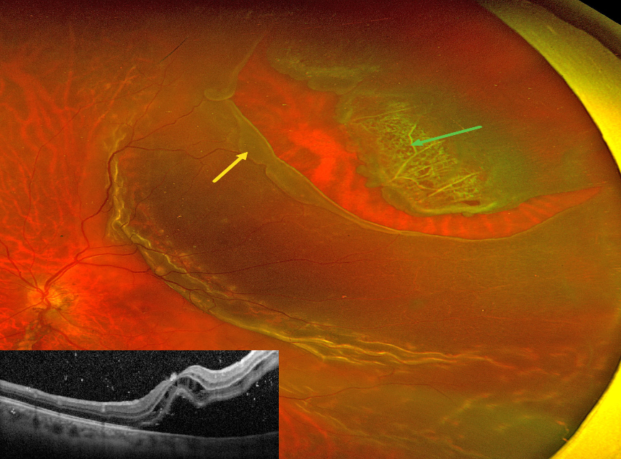
This 46YO male presented with recent vision loss in his left eye. Vision was counting fingers.
Optos color RG imaging shows a superotemporal macula-off rhegmatogenous retinal detachment (RRD). The anterior flap of the causative tear encompasses an entire lattice lesion. The posterior edge of the tear has a posteriorly-rolled edge, indicating early Grade B proliferative vitreoretinopathy (The Retina Society Terminology Committee, Ophthalmology 1983;90:121-125).
OCT shows cystic retinal edema mostly in the outer nuclear layer, along with hyperreflective vitreous and subretinal cells. The patient was scheduled for emergent vitrectomy.



