

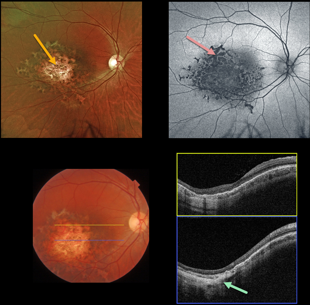
This healthy 32YOF was referred for an asymptomatic lesion in her right macula. Vision was 20/30 OU, and her left fundus was normal.
Optos color imaging shows an atrophic, variably pigmented macular colobomatous lesion with a surrounding rim of scalloped pigmentation.
Fundus autofluorescence (FAF) shows variable, mostly hypo-FAF, indicating RPE dysfunction/loss.
Swept source OCT scanning shows outer retinal/RPE thinning with a colobomatous/staphylomatous outpouching.
Learning Points:
Retinal pigment epithelium dysgenesis (RPED) was first described by Cohen et al (Arch Ophthalmol 2002;120:512-516) as a unilateral, idiopathic lesion affecting the RPE and outer retina. These rare lesions have a pathognomonic scalloped margin with associated RPE atrophy and fibrosis.
Although initially described as a unilateral condition, it can also appear bilaterally (Renz et al, Arch Ophthalmol 2012;130:1341-1340). The lesions can enlarge with time and also carry a small risk for secondary macular neovascularization (Shimoyama et al, Case Rep Ophthalmol 2014;5:34-37) and presumed RPE tumor (Gal-Or et al, Retinal Cases & Brief Reports 2019;13:121-126).
To the best of our knowledge, our patient’s lesion is unique by the colobomatous appearance which has not been previously described.



