

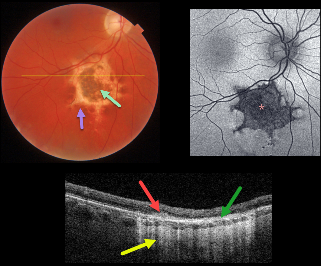
This asymptomatic 34YO male was referred for an asymptomatic lesion just inferior to the right macula. The 2-disc-diameter lesion is hyperpigmented centrally with a scalloped hypopigmented border.
Optos fundus autofluorescence (FAF) shows diffuse hypo-FAF of the lesion due to a lack of RPE lipofuscin from the RPE and outer retinal atrophy.
Triton swept-source OCT through this lesion shows outer retinal hyperreflectivity within the area of hyperpigmentation, disorganized atrophic outer retinal layers and RPE, and hyperreflectivity of the choroid and sclera due to increased light transmittance through the outer retinal and RPE atrophy.
The patient will be monitored yearly for any changes.
Learning Points:
Retinal pigment epithelium dysgenesis (RPED) was first described by Cohen et al (Arch Ophthalmol 2002;120:512-516) as a unilateral, idiopathic lesion affecting the RPE and outer retina. These rare lesions have a pathognomonic scalloped margin with associated RPE atrophy and fibrosis.
Although initially described as a unilateral condition, it can also appear bilaterally (Renz et al, Arch Ophthalmol 2012;130:1341-1340). The lesions can enlarge over time and also carry a small risk of secondary macular neovascularization (Shimoyama et al., Case Rep Ophthalmol 2014;5:34-37) and a presumed RPE tumor (Gal-Or et al., Retinal Cases & Brief Reports 2019;13:121-126).



