

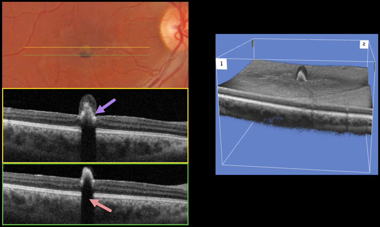
This 26YO male was referred for a pigmented lesion in his right eye. He has a lifelong history of poor vision in this eye and denied any new visual symptoms. Vision was 20/200 OD and 20/20 in his normal left eye.
Triton color imaging shows an inner retinal nodular hyperpigmented lesion just inferior to the macular center. Swept-source OCT shows marked hyperreflectivity from the lesion with posterior shadowing. The superior portion of the lesion is relatively depigmented and shows less OCT hyperreflectivity. 3D OCT reconstruction shows another view of this lesion.
Learning Points:
RPE hamartoma was originally described by Gass as a “focal, nodular, jet-black lesion” that can extend through the internal limiting membrane (Eye 1989;3:1-18). The lesion is markedly hyperreflective on OCT with complete posterior shadowing, although deeper intralesional retinal structures can be visualized with enhanced-depth imaging (Takahashi et al, Retinal Cases & Brief Reports 2013;7:179-183). Vision can be normal or decreased if the lesion involves the central macula, as in our patient. The lesions show no underlying RPE or choroidal changes, remain unchanged with observation, and require no treatment.



