

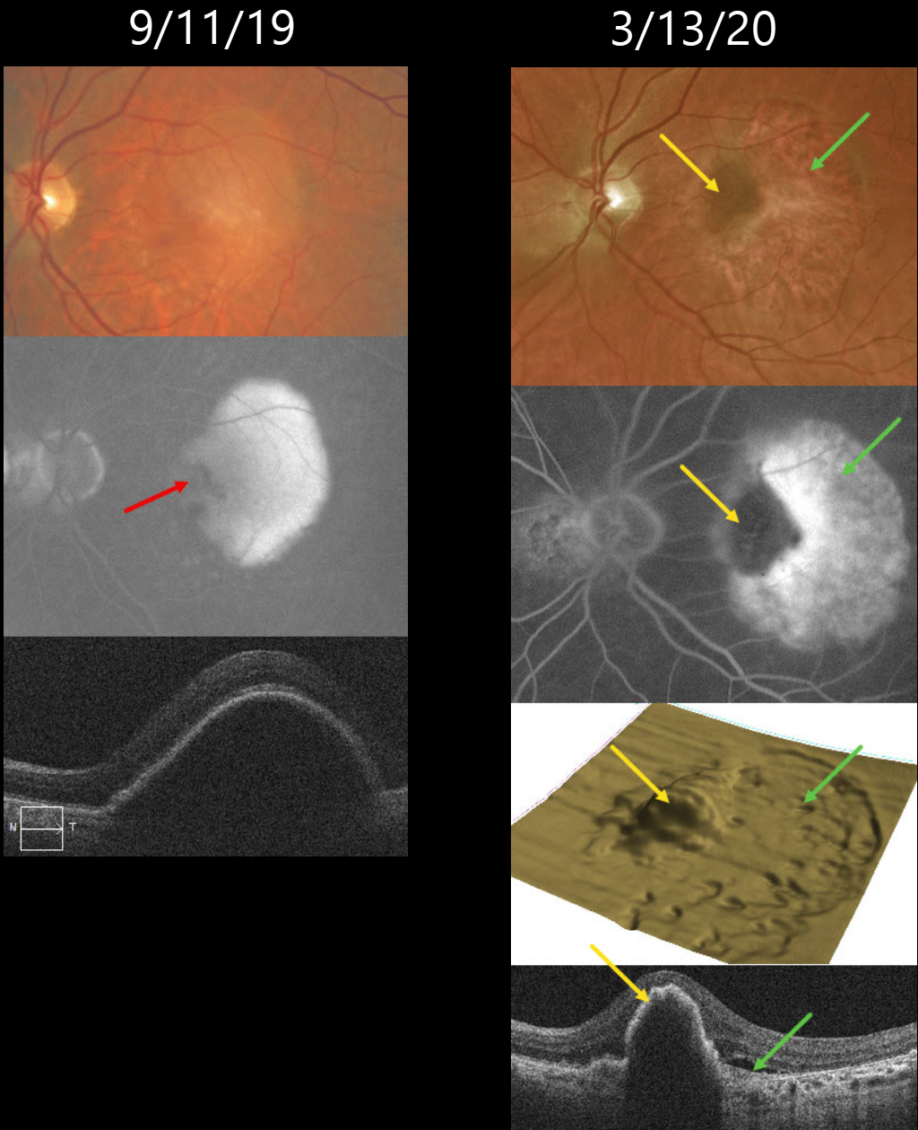
This 72YO male was followed for many years with stable intermediate AMD. On 9/11/19, he presented with a new symptomatic retinal pigment epithelial detachment (PED) with 20/30 vision. Fluorescein angiography shows an uneven-filling PED with a nasal hypofluorescent notch. A large PED is confirmed on OCT scanning.
He initially did well with monthly Eylea injections. While on treat-and-extend therapy, he presented on 3/13/20 without new visual symptoms, although his vision had decreased to 20/200.
Optos color RG imaging shows a new, large temporal RPE tear. On multimodal imaging, the bed of the tear is depigmented on color imaging, hyperfluorescent on fluorescein angiography (window defect), and shows a crater on the 3D OCT RPE map, and shows bare Bruch’s membrane on B-scan.
The retracted edge of the tear is hyperpigmented on color imaging, hypofluorescent on fluorescein angiography (blockage), an elevated mound on the 3D OCT RPE map, and a rippled, elevated PED on B-scan.
We subsequently decided on PRN therapy, with the last injection given on 3/12/21. When last examined, vision was stable at 20/400 with a stable dry lesion.
Learning Points:
A tear in the RPE is a fairly common occurrence in PED associated with type 1 macular neovascularization (MNV) located below the RPE. The neovascularization can spontaneously contract or involute following treatment. It then puts traction on the overlying RPE, which can then rip at the opposite edge of the PED.
Like a cheap window blind, the torn RPE retracts and scrolls towards the MNV. This leaves a depigmented region where the RPE was and is now absent, along with a pigmented, relatively straight subretinal lesion from the scrolled RPE edge.
Originally described by Gass (Retina 1984;4:205-220), a notched PED is a sign of an occult MNV. The notch represents a type 1 MNV, which tethers the overlying RPE from the surrounding more highly elevated PED. Hydrostatic leakage from the MNV may contribute to the larger PED.



