

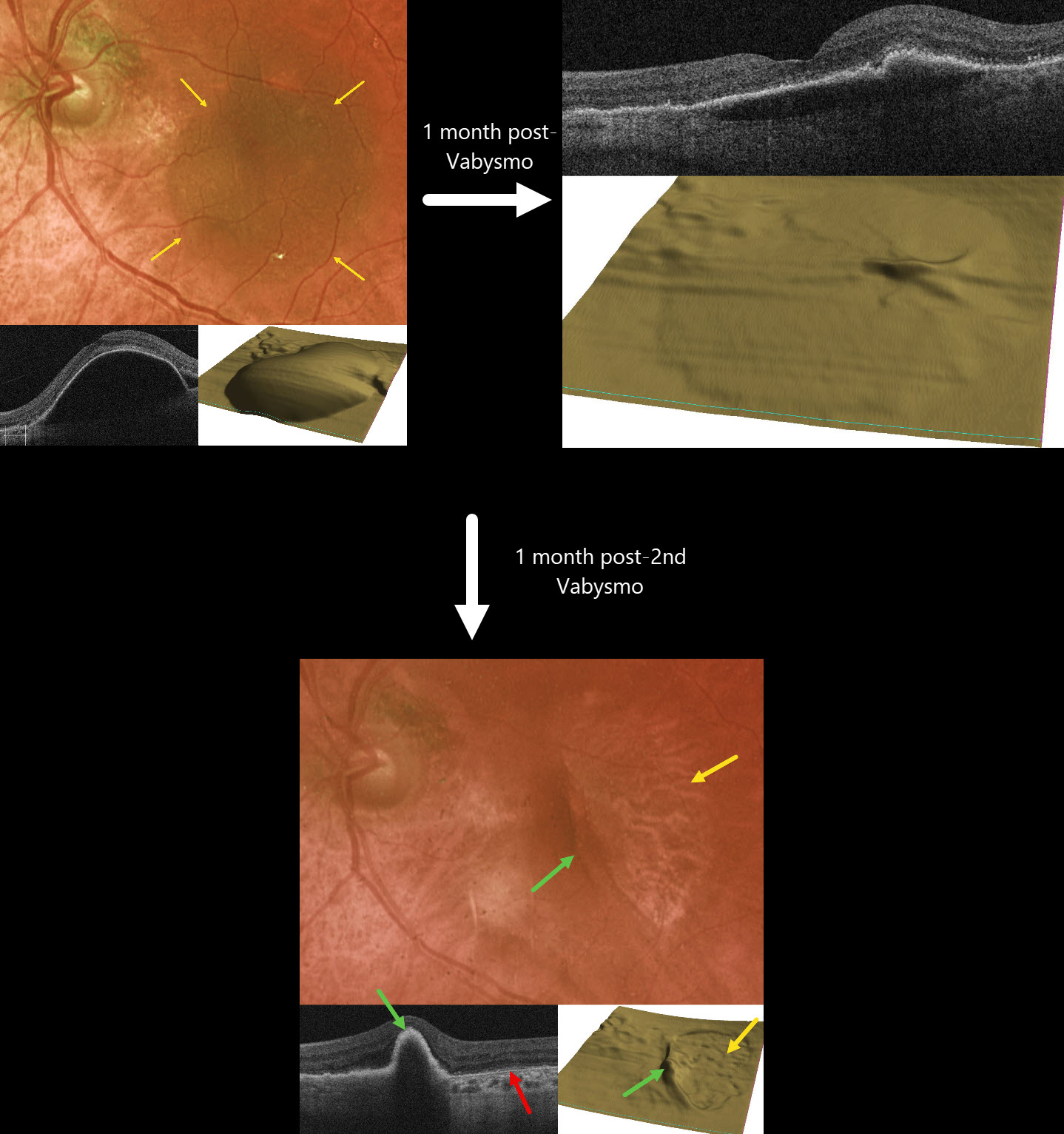
This 77YO female presented with recent vision loss in her left eye from new neovascular AMD. Vision was 20/400.
Optos color RG imaging shows a large pigmented retinal pigment epithelial detachment (PED) occupying most of the left macula. OCT scanning, B-scan, and RPE map confirm this large PED. Intravitreal Vabysmo was injected, and one month later, vision improved to 20/40. The PED nearly flattened.
Another Vabysmo injection was given, and one month later, she complained of some new vision loss. Vision was 20/80. A new RPE tear is noted, with bare RPE temporally and the retracted RPE nasally. Bare Bruch membrane is seen within the bed of the torn RPE. Vabysmo was injected, and treat-and-extend was begun.
Learning Points:
A tear of the RPE is a fairly common finding in RPE detachment (PED) associated with type 1 macular neovascularization (MNV, located below the RPE). Neovascularization can spontaneously contract or involute following treatment. It then puts traction on the overlying RPE, which can then rip at the opposite edge of the PED. Like a cheap window blind, the torn RPE retracts and scrolls towards the MNV. This leaves a depigmented region where the RPE was and is now absent, along with a pigmented, relatively straight subretinal lesion from the scrolled RPE edge. Anti-VEGF therapy can safely be continued following an RPE tear, with long-term visual outcome more closely related to the patient’s response to therapy than to the tear itself (Ophthalmology 2018;125:236-244), provided the tear is outside the foveal center.



