

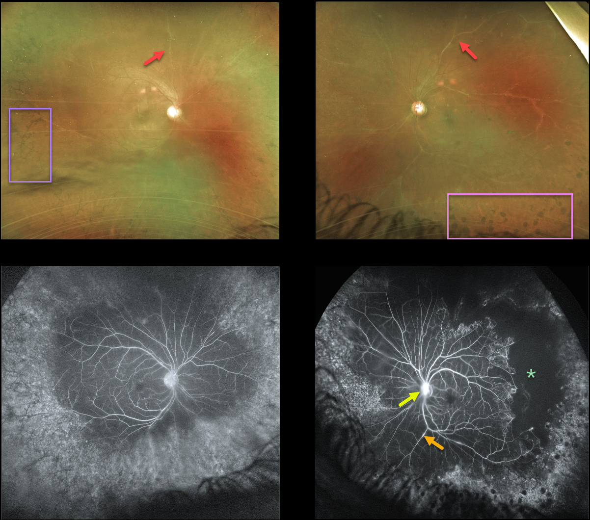
This 43YO male has a long history of panuveitis from sarcoidosis, and was previously seen 4 years earlier before being lost to follow-up.
He presented recently with a few weeks of symptomatic panuveitis. Vision was 20/100 OD and 20/200 OS with active granulomatous anterior uveitis.
Optos imaging shows a hazy view of each fundus, with diffusely white retinal vessels. Inferior vitreous puff balls were noted clinically but not imaged on Optos.
Fluorescein angiography shows focal areas of vascular staining/leakage, severe peripheral ischemia, and late optic nerve staining/leakage (especially OS). There is encircling peripheral intraretinal pigment migration OD with apparent peripheral scatter laser scarring OS.
He is being treated with topical steroids since the majority of active inflammation is anterior.
Further scatter laser to the untreated ischemic retina will be recommended if posterior segment neovascularization develops.



