

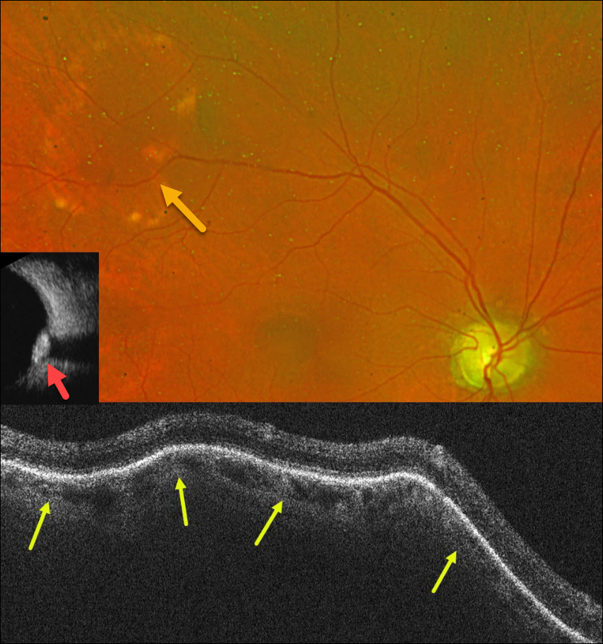
This 76YO male was referred for an asymptomatic lesion in his right fundus. Vision was 20/30.
Optos color RG imaging shows a lumpy, elevated subretinal yellow-white nodular lesion in the superotemporal midperiphery.
OCT scanning through this lesion shows marked lumpy scleral elevation with distortion of the overlying choroid and retina. This lesion was markedly hyperreflective on B-scan ultrasonography, indicating calcification. The left fundus was normal (not shown).
Learning Points:
Sclerochoroidal calcification is a relatively uncommon and benign finding in the elderly population. These yellowish lesions are slightly lumpy and elevated, and usually appear in the mid superior/inferior temporal/nasal quadrants. The lesions are highly reflective on B-scan ultrasonography and are also visible on CT scanning. They require no treatment and are not associated with vision loss.
However, they may be associated with systemic hypercalcemic conditions such as hyperparathyroidism and pseudohypoparathyroidism. Shields et al therefore recommended that all such patients be evaluated for underlying systemic calcium disorders (Retina 2015;35:547-554). Our patient’s bloodwork was fortunately negative.



