

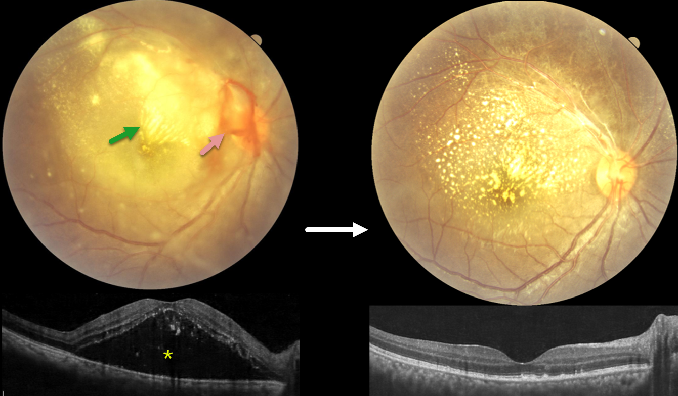
This 28YO female presented with 4 days of blurred vision in her right eye. For the past 2 weeks, she has complained of generalized weakness, malaise, and body pain. Vision was counting fingers OD and 20/20 in her normal OS.
Fundus photography shows variable, dense subretinal lipid, particularly confluent in the superonasal macula. There is some preretinal blood around the nerve, and the retina is somewhat yellow and opaque temporally and inferiorly. Patches of inner retinal opacification are noted outside the macula superiorly. OCT shows hyperreflective dots from lipid exudate in the outer retina, with a large serous detachment. Laboratory testing, including CBC, ESR, syphilis, TB, HIV, TORCH profile, and chest X-ray, was negative. We started a 1-week tapering course of oral prednisone 40mg and doxycycline 100mg BID. Three days after our initial examination, she mentioned that she had 2 weeks of mild intermittent fever. Weil-Felix testing was positive, and she was subsequently diagnosed with scrub typhus and asked to continue the doxycycline for a full 2-week course.
The clinical picture gradually improved, and about 6 weeks following presentation, vision improved to 20/40 despite significant residual lipid. OCT scanning through the macular center was mostly normalized. We continue to follow her closely.
Learning Points:
Rickettsial infections cause posterior segment involvement in a substantial proportion of patients, often asymptomatic despite significant fundoscopic findings. The most common rickettsial organisms causing retinal disease include Rickettsia rickettsii (Rocky Mountain spotted fever), Rickettsia conorii (Mediterranean spotted fever), and Rickettsia typhi (murine typhus).
Scrub typhus, caused by Orientia tsutsugamushi and transmitted by a larval mite (chigger) bite, has ocular involvement in about 20% of patients. Retinal findings include retinal hemorrhages, branch retinal vein occlusion, retinal vasculitis/retinitis, and papillitis (Ganekal et al, Ind J Ophthalmol 2021;69:1167-1171).
The ocular and systemic findings of vasculitis and perivasculitis result from both direct endothelial cell destruction by the organism and exaggerated immune responses. Doxycycline is the drug of choice; treatment should last at least 7 days and continue for at least 3 days after fever has subsided and clinical improvement is evident. Visual prognosis is generally favorable with appropriate treatment.



