

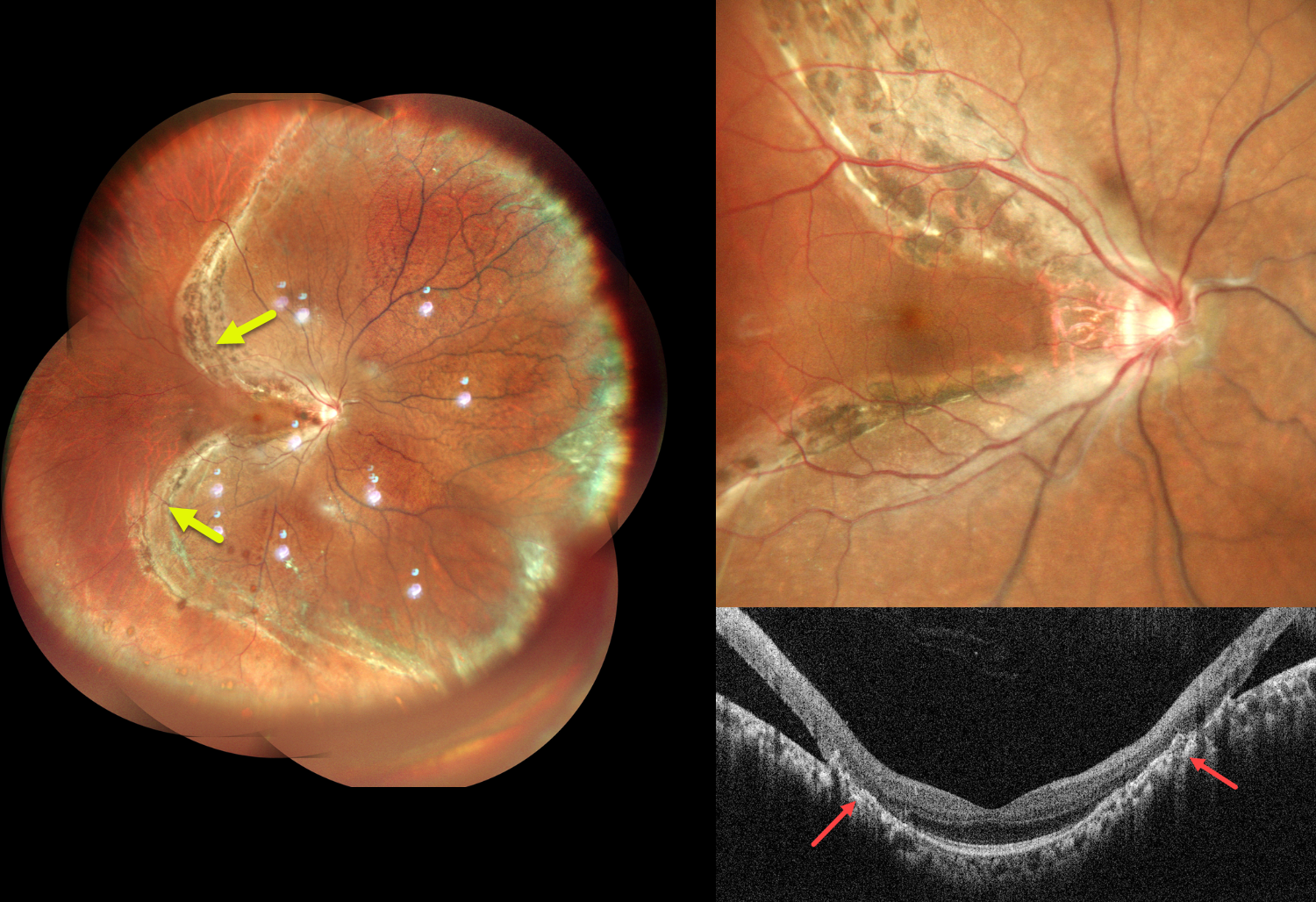
This healthy 31YO male presented with blurred vision in his right eye for 2 years. Best-corrected visual acuity was 20/20 OU.
Pseudocolor SLO imaging shows a chronic rhegmatogenous retinal detachment (RRD) involving the superior, nasal, and inferior quadrants, sparing the central and temporal macula. A prominent pigmented demarcation encircles the RRD. Vertical OCT scanning shows a normal macular center with broad chorioretinal scarring superiorly and inferiorly bordering the attached and detached retina.
Learning Points:
Acute RRDs are virtually always symptomatic, requiring emergent surgery. However, asymptomatic detachments often have a low risk of progression and can usually be safely observed (Cohen et al, AJO 2005;139;777-779; Sengillo et al, Ophthalmology Retina 2023;7:215-220) or demarcated (Vrabec and Baumal, Ophthalmology 2000;107:1063-1067).
In the present case, although the retinal detachment was symptomatic at onset, it was not diagnosed until several years later. The heavy demarcation line indicates chronicity with likely stability. Given the preserved visual acuity, macular sparing, and chronic nature of the detachment, close observation was recommended.



