

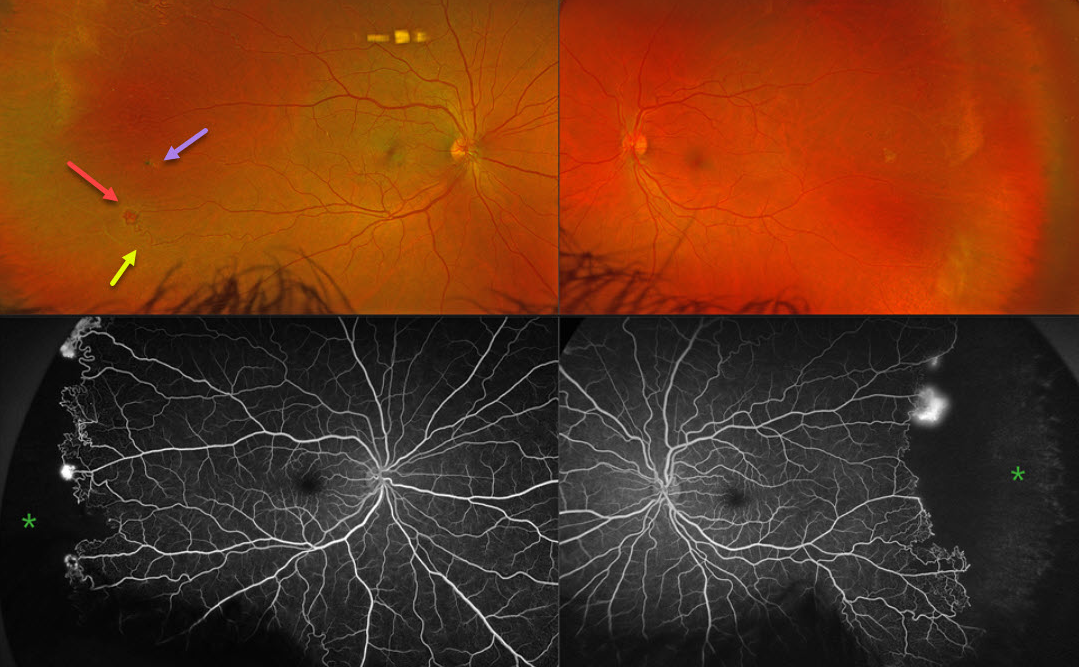
This 43YO male was referred for asymptomatic retinal findings. He has a known history of sickle cell disease. Vision was 20/20 OU.
Optos color RG imaging shows bilateral peripheral temporal and featureless retinas with white vessels. There is a small temporal pigmented chorioretinal scar OD, indicating a possible black sunburst lesion. The retinal vascular bed is remodeled along the border of perfused and ischemic retina with retinal arteriovenous shunting and neovascularization. The complete temporal nonperfusion with bilateral neovascularization is confirmed on fluorescein angiography.
Learning Points:
Patients with sickle cell disease develop symptoms throughout their bodies as stiffened, sickled red blood cells block capillary beds. In the eye, this can cause proliferative sickle cell retinopathy (PSR) with sea-fan-shaped areas of peripheral retinal neovascularization developing at the junction of perfused and ischemic retina. Complications include vitreous hemorrhage and tractional retinal detachment. Although patients with SS disease tend to have more systemic complications, SC disease is usually associated with more severe ocular findings.
Treatment with sectoral (Farber et al Arch Ophthalmology 1991;109:363-367) or circumferential (Kimmel et al Ophthalmology 1986;93:1429-1434) scatter laser photocoagulation is somewhat controversial since, unlike more typical proliferative retinopathies (ie from diabetes and branch retinal vein occlusions), these new vessels tend to auto-infarct with a good natural history for untreated disease (Downes et al Ophthalmology 2005;112:1869-1875). We elected to closely follow him for now with observation.



