

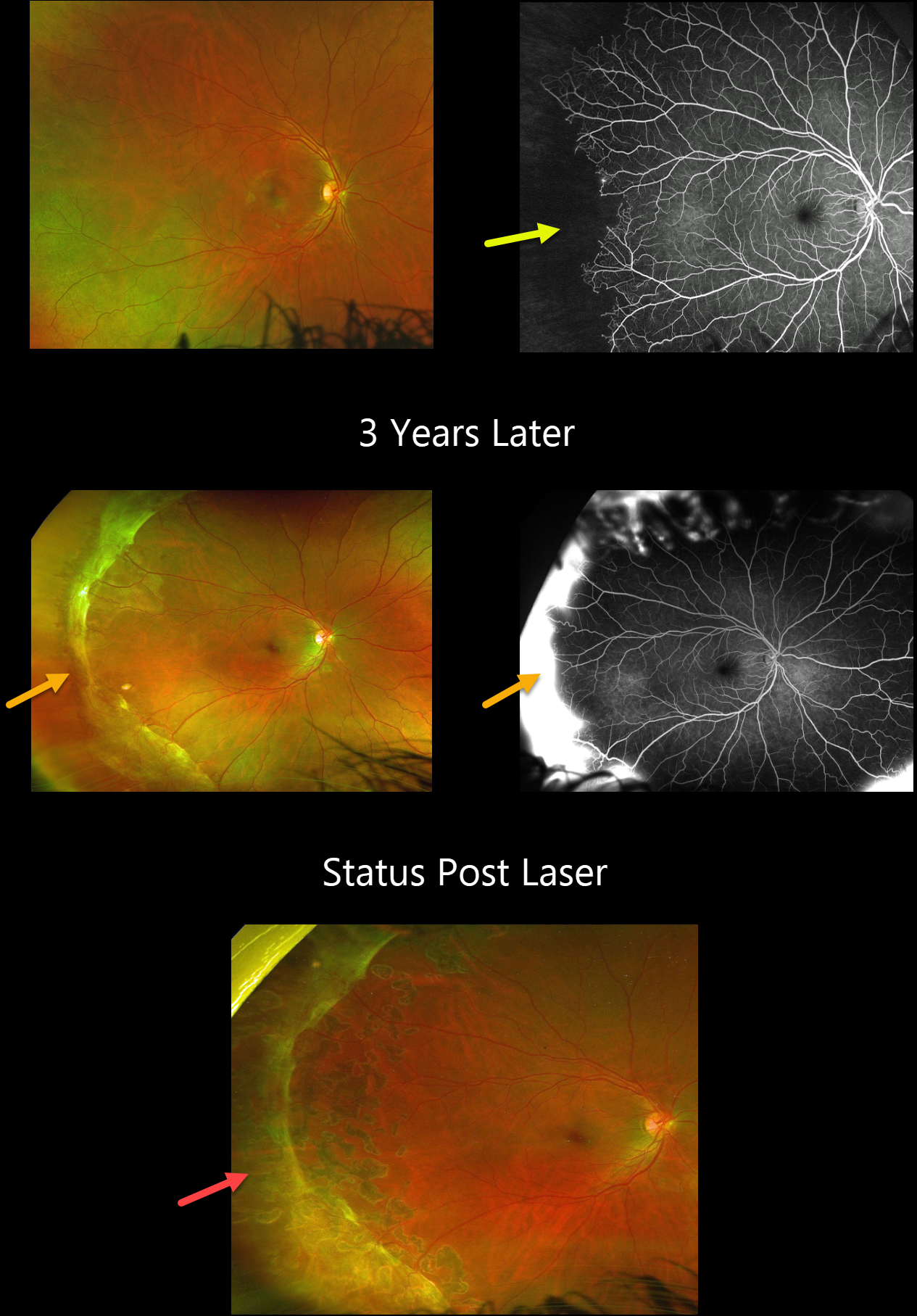
This 19YO male has a known history of sickle disease (Hb SC). On 7/30/20, vision was 20/25 OD.
Optos color imaging shows temporal vascular whitening, and angiography shows marked temporal ischemia.
He was lost to follow-up until 1/10/23 when he presented with hand motions vision from a vitreous hemorrhage OS (not shown). Although vision was still 20/20 OD, a broad band of fibrosed neovascularization was noted in the periphery. A peripheral scatter laser was applied to the right eye at the time of OS vitrectomy, and 6 weeks later, all neovascularization had regressed.
Learning Points:
Patients with sickle cell disease develop symptoms throughout their bodies as stiff, sickled red blood cells block capillary beds. In the eye, this can cause proliferative sickle cell retinopathy with sea-fan shaped areas of peripheral retinal neovascularization developing at the junction of perfused and ischemic retina. Complications include vitreous hemorrhage and traction retinal detachment. Although patients with SS disease tend to have more systemic complications, SC disease is usually associated with more severe ocular findings.



