

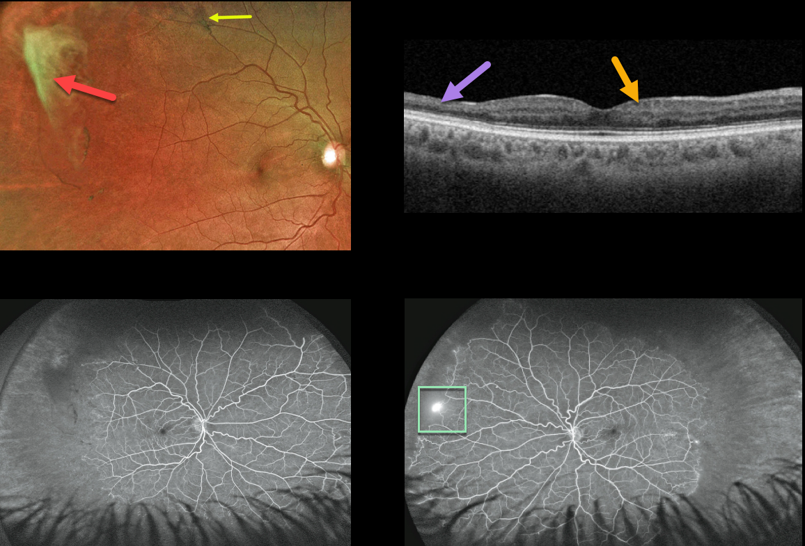
This 39YO African American male presented with asymptomatic proliferative sickle cell retinopathy with spontaneously avulsed bilateral superotemporal peripheral fibrosed retinal neovascularization (sea fans). A small midperipheral black sunburst lesion is seen superotemporally in his right eye.
Swept-source OCT shows variable inner retinal thinning and disorganization of the retinal inner layers (DRIL).
Fluorescein angiography shows a patch of retinal neovascularization OS at the junction of perfused and non-perfused retina.
Since these lesions often auto-infarct, as they did in our patient, we elected to observe the small area of neovascularization in his right eye. He is also at low risk for a vitreous hemorrhage or traction detachment since the vitreous is already detached in this area.
Learning Points:
Patients with sickle cell disease develop symptoms throughout their bodies as stiff, sickled red blood cells block capillary beds. In the eye, this can cause proliferative sickle cell retinopathy (PSR) with sea-fan-shaped areas of peripheral retinal neovascularization developing at the junction of perfused and ischemic retina.
The variable macular thinning noted on OCT is caused by microinfarcts to the retinal microcirculation. Enlargement of the foveal avascular zone is a common OCT angiographic finding (Fares, AJO 2021;224:7-17).
The black sunburst lesions are thought to occur from intraretinal RPE migration in response to photoreceptor damage from intraretinal and subretinal hemorrhage (see Ausdourian et al, BJO 1975;59:710-716).



