

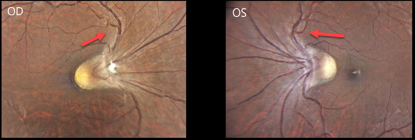
This healthy 38YO highly myopic female presented with 6 months of decreased vision in her right eye. Her current refraction was -15.00 D OD and -11.00 -2.25 × 10° OS. Vision was 20/200 OD and 20/25 OS.
Color photography shows the retinal vessels emerge from slightly tilted discs to initially course nasally and superiorly before turning temporally toward the macula. The macular center is normally located about 2-disc diameters temporal to the nerve. The right fovea cannot be visualized and likely lies just temporal to the edge of the nerve, and about one-half disc diameter temporal to the left nerve.
Learning Points:
Situs inversus occurs in 5% of normal eyes and 70% of eyes with tilted discs (Witmer et al, Surv Ophthalmology 2010;55:403-428) and presents with the retinal vessels exiting the optic disc nasally. Vision is usually normal unless other pathology is present.
Despite the abnormal macular architecture, our patient reportedly had normal vision until recently, and was found to have a cataract, which was consistent with her acuity. Cataract surgery was recommended, and vision improved to 20/25 postoperatively.



