

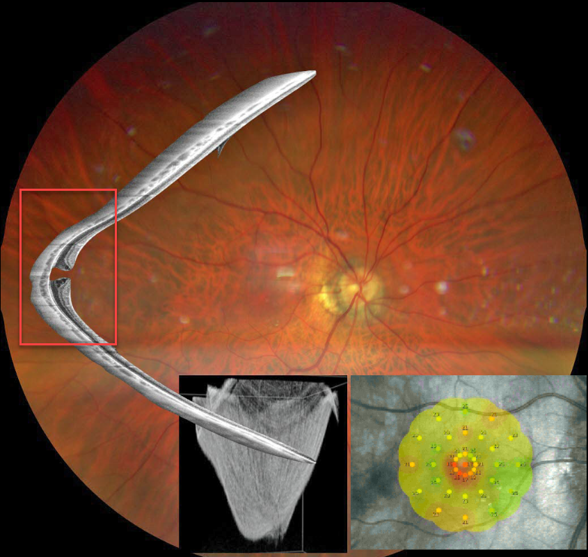
This Eyecare Clinic (in Brescia, Italy) case was submitted by Barbara Parolini, Veronika Matello, Giulia Freschi, and Roberta Penzani.
This 48YO highly myopic male presented for a sudden drop in vision. Vision was 20/100 OD and 20/20 in OS.
Color photography shows a blunted macular reflex. Widefield OCT shows a conical-shaped staphyloma of the entire posterior pole with a full-thickness macular hole. Microperimetry shows a decreased but not absent foveal sensitivity.
According to the Myopic Traction Maculopathy Staging System (Parolini et al, Eur J Ophthalmol 2021;31:1299-1312), this eye is in Stage 1c. The left eye showed a similar staphyloma but without a macular hole (not shown). The patient declined surgery.
Learning Points:
A macular staphyloma consists of a concave posterior bowing of the eye wall in patients with high myopia. These myopic patients also have a markedly thinned choroid throughout the posterior pole.
Due to stretching of the posterior retinal structures, these eyes are at increased risk of numerous findings, including macular schisis/traction and macular neovascularization.



