

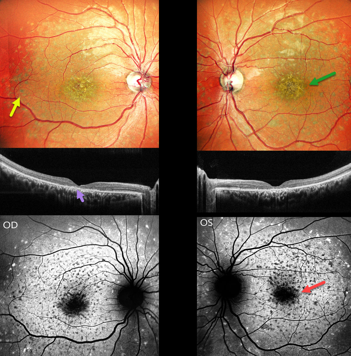
This 26YO male presented with 3 years of bilateral decreased vision along with night vision difficulties since childhood. Family history was negative. Vision was counting fingers at 3 meters OU.
Pseudocolor SLO imaging shows a symmetrical ‘beaten-bronze’ sheen to each central macula with surrounding yellowish pigmented flecks. OCT shows central loss of the photoreceptor layers with hyperreflective deposits above the RPE. Green fundus autofluorescence (FAF) shows foveal hypo-FAF. The clinical flecks are hypo-FAF centrally and hyper-FAF more peripherally. There is relative peripapillary sparing.
Learning Points:
Stargardt disease is an autosomal recessive disorder caused by a mutation in the ABCA4 gene located on chromosome 1. The ABCA4 transmembrane protein localizes to the photoreceptor outer segments and is involved in the recycling of 11-cis-retinal. Mutations of this gene cause the accumulation of lipofuscin in the RPE, which eventually leads to photoreceptor and RPE degeneration. Other ABCA4 disorders include fundus flavimaculatus, cone-rod dystrophy, retinitis pigmentosa, and age-related macular degeneration.
Historically, Stargardt disease was used to describe patients with macular involvement only, and fundus flavimaculatus for those with flecks extending more peripherally. Both are now considered different retinal phenotypes within the ABCA4 spectrum.



