

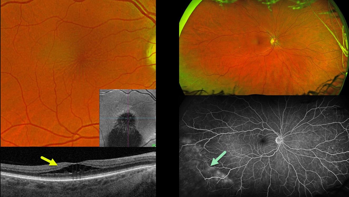
This 58YO female presented with 6 months of blurred vision in her right eye. Vision was 20/40 OD and 20/25 OS.
Optos color RG imaging of her right eye shows a stellate pattern of foveal schisis, which is confirmed on Triton swept-source B-scan and en face OCT. The macular appearance and OCT in her left eye were normal (not shown). Inferotemporal retinoschisis is noted OU peripherally. The peripheral schisis shows late leakage angiographically. Genetic testing was heterozygous for a pathogenic USH2A mutation.
Learning Points:
A new classification of stellate nonhereditary idiopathic foveomacular retinoschisis (SNIFR) was initially described by Ober et al (Ophthalmology 2014;121:1406-1413). Unlike X-linked schisis, which, in our experience, mostly affects the inner and outer nuclear layers bilaterally, SNIFR causes splitting of the outer plexiform layer, usually in just one eye. Many eyes will have peripheral retinoschisis, even when the macula is normal (Bloch et al, Retina 2021;41:2361-



