

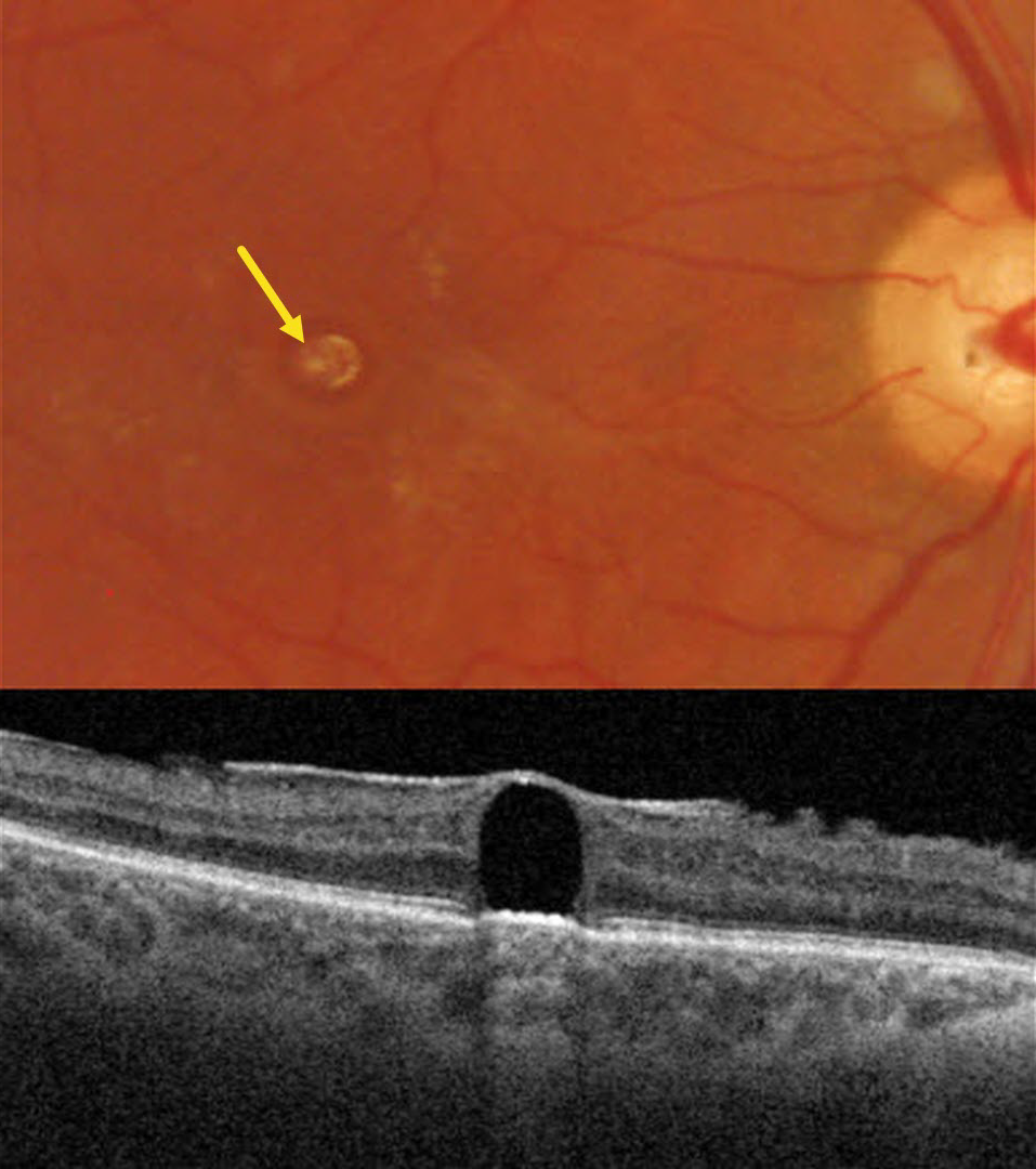
This 33YO male presented with stable vision following prior vitrectomy for a rhegmatogenous retinal detachment (RRD) in his right eye. Vision was 20/400.
Triton color imaging shows a round yellowish subfoveal lesion. Swept-source OCT shows a hyporeflective subretinal lesion compressing the overlying neurosensory retina into a thin inner retinal hyperreflective line. The underlying RPE band is slightly anteriorly displaced, with hyperreflective choroidal shadowing. A mild epiretinal membrane is also noted.
Learning Points:
Perfluorooctane (PFO) is a liquid heavier than water used intraoperatively for complex retinal detachments, including proliferative vitreoretinopathy and giant retinal tears. The PFO is removed during the fluid-air exchange. Rarely, PFO droplets can develop intraoperatively and migrate subretinally. These were much more common from intraocular turbulence prior to the introduction of modern valved cannulas.
Subretinal PFO bubbles have a characteristic appearance on OCT, including an acute angle between the bubble and the RPE, thinned, ill-defined overlying retinal layers, elevation of the underlying RPE band, and hyperreflective choroidal shadowing (Figueroa and Contreras, Retina 2012;32:2177-2178). The subretinal PFO can be removed using a variety of surgical maneuvers, although we recommend observation given its chronicity and absence of new visual complaints.



