

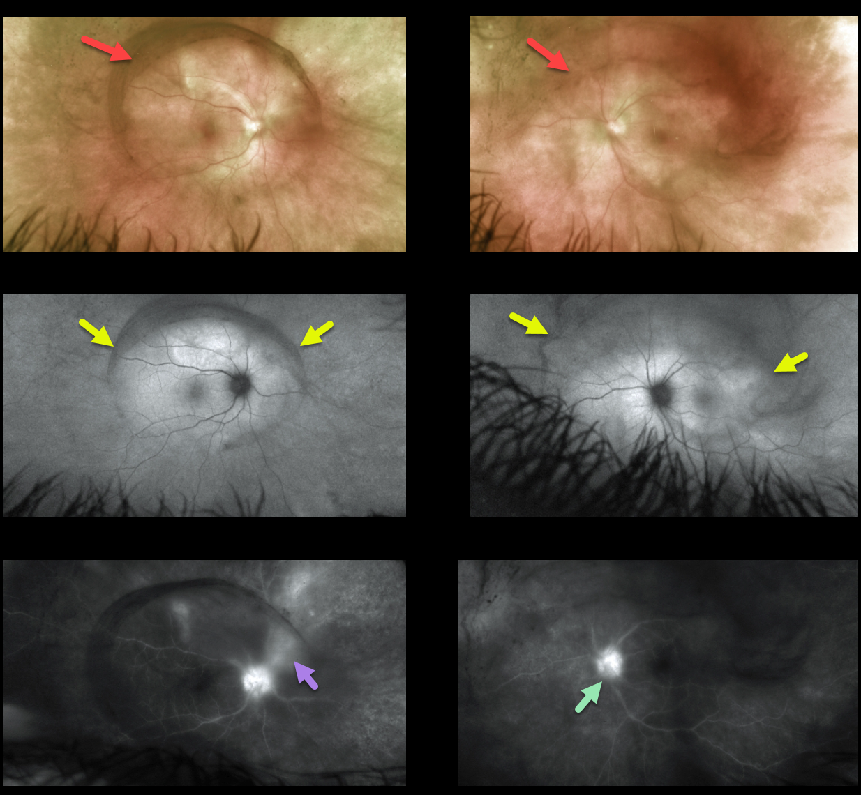
This 49YO female presented with a 1-month history of bilateral vision loss and eye pain. Vision was 20/200 OU. There was a mild-moderate, bilateral nongranulomatous anterior uveitis.
Vitreous debris was centered over each posterior pole, which was grossly normal.
Fundus autofluorescence (FAF) shows an oval region of placoid hyper-FAF in each posterior pole. Fluorescein angiography shows bilateral nerve leakage and vasculitis.
RPR and treponema pallidum antibody testing were positive, and she was immediately started on intravenous penicillin.
Learning Points:
Ocular manifestations of syphilis vary widely and include virtually all variations of anterior and posterior segment inflammation, including a recently described presentation as the multiple evanescent white dot syndrome phenotype (see Russel et al Int Ophthalmol 2020;40:627-738). Hence, the reason it’s been dubbed the great imitator or masquerader.
One of the more common presentations is placoid chorioretinitis, which was evident on FAF imaging in our patient.
Patients also need to be evaluated for HIV since syphilis and HIV infections often coexist. Fortunately, our patient was HIV negative.



