

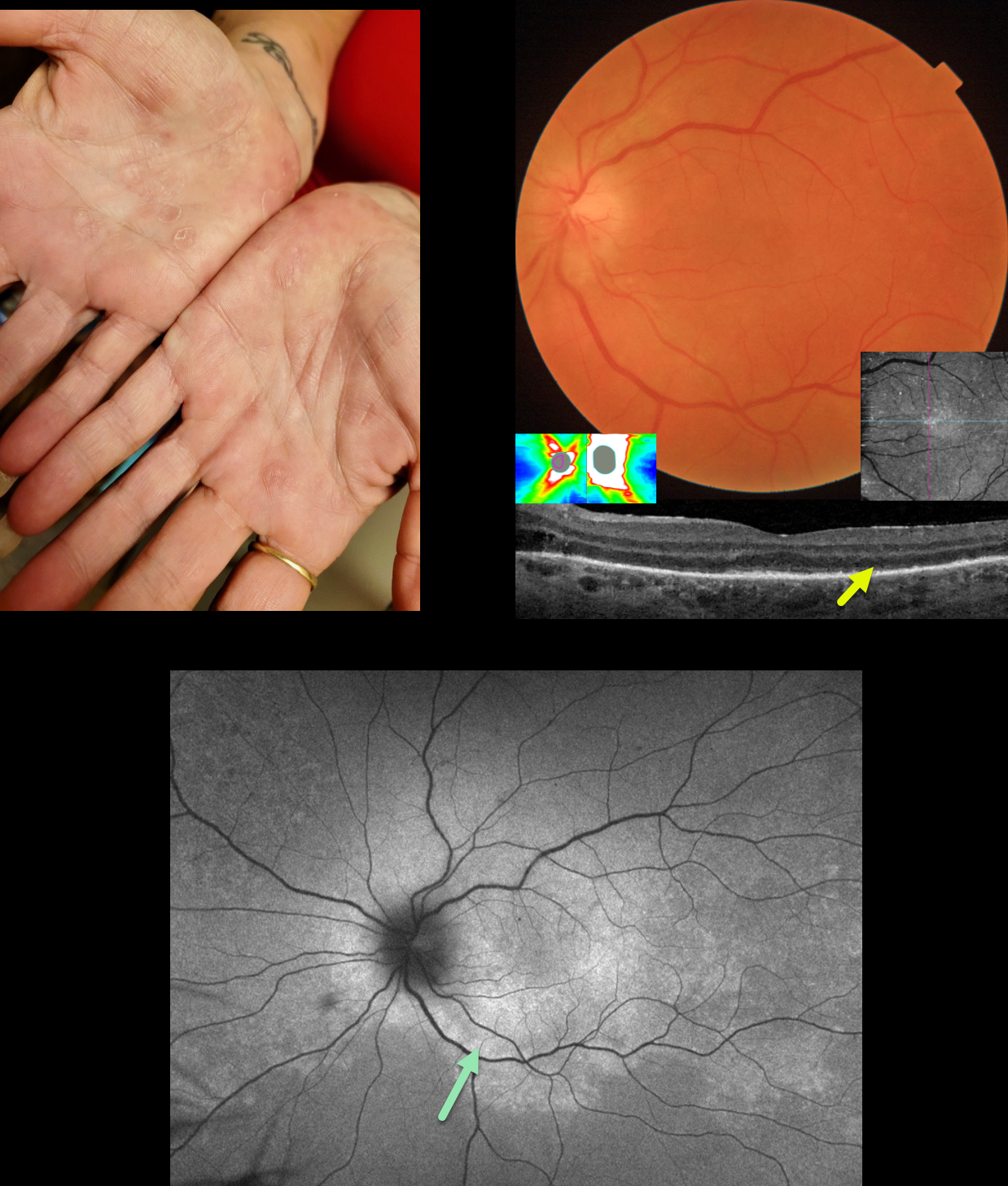
This 51YO female presented with one month of flashes and vision loss in her left eye. Vision was 20/20 in her normal OD and counting fingers OS.
When asked about any rashes, she showed us her palms. What’s your diagnosis??
Triton color imaging of the left posterior pole shows mild optic nerve swelling, with some subtle subretinal whitish pigmentary changes. The nerve swelling was confirmed on OCT. Swept-source OCT shows variable loss of the outer retinal bands and ellipsoid zone with hyperreflective material above the RPE. En face imaging of the outer retina shows this hyperreflective material as numerous bright dots.
Fundus autofluorescence (FAF) shows confluent macular and peripapillary hyper-FAF with more patchy areas of hyper-FAF extending peripherally. Laboratory testing was positive for syphilis and negative for HIV. She was referred to an infectious disease specialist for systemic penicillin treatment and was subsequently lost to follow-up.
This case was submitted by Mattie Adams.
Learning Points:
Ocular manifestations of syphilis vary widely and include virtually all variations of anterior and posterior segment inflammation, including a recently described presentation as the multiple evanescent white dot syndrome phenotype (Russel et al Int Ophthalmol 2020;40:627-738). Hence, the reason it’s been dubbed the great imitator or masquerader. One of the more common presentations is placoid chorioretinitis (Eandi et al, Retina 2012;32:1915-1941), which was evident in our patient’s multimodal imaging.
The rates of syphilis in the US have steadily climbed since 2000, and according to the CDC, its incidence rose nearly 80% between 2018 and 2022 (https://www.cdc.gov/std/statistics/2022/default.htm). Syphilis is prevalent in men having sex with men, among heterosexuals, and in both sexes. These numbers are reflected in how often we are now finding and diagnosing syphilis in our practice. We must always remember that as eye doctors, we are uniquely privileged to not only treat eye problems, but also diagnose systemic diseases. In our case, the painless palmar rash and macular placoid findings were both pathognomonic and led to a rapid diagnosis with referral for treatment.



