

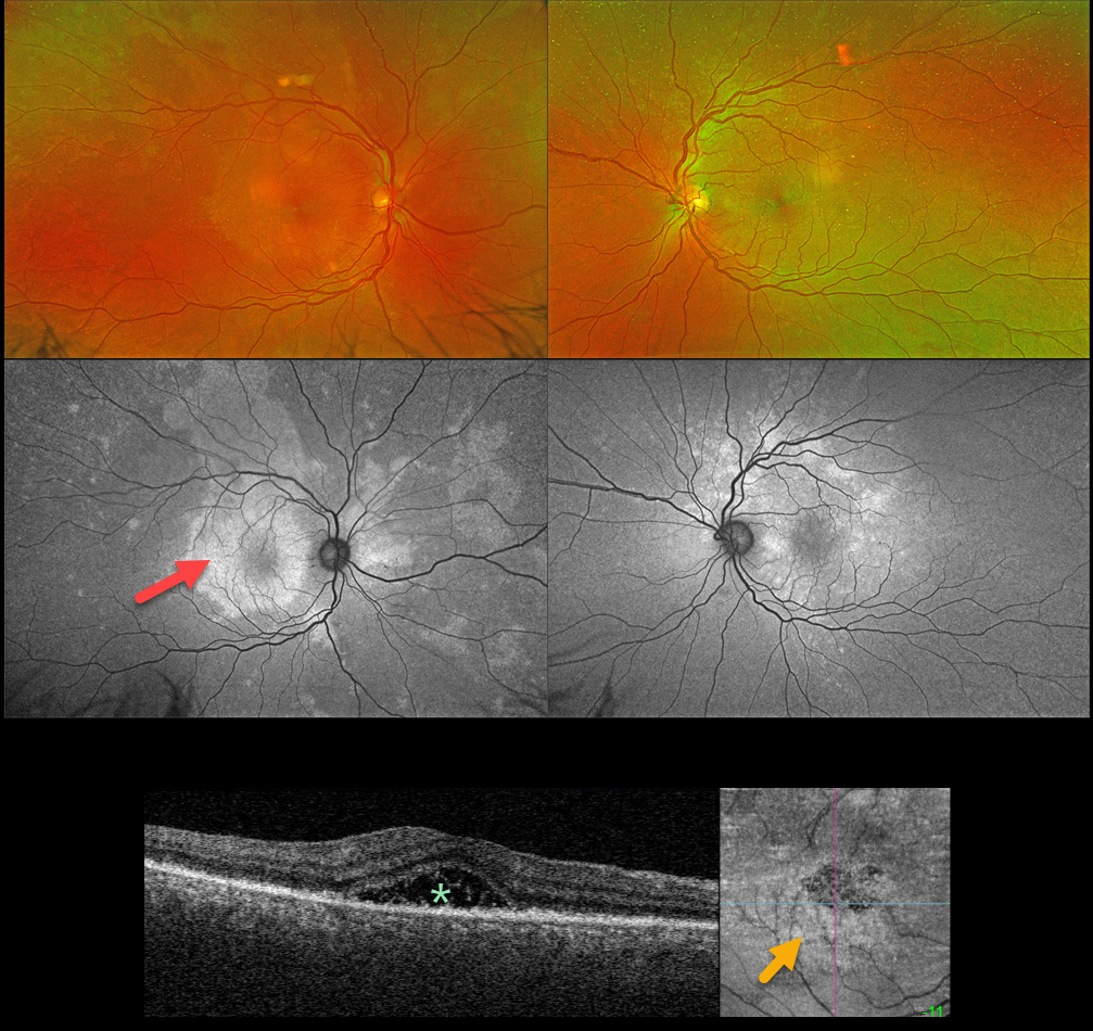
This 52YO male presented with a one-week history of blurred central vision in his right eye. There was no past medical history. Vision was 20/200 OD and 20/25 OS. Anterior segments were normal.
An oval, whitish submacular lesion is noted OD. Fundus autofluorescence (FAF) reveals much more extensive findings than those noted funduscopically. In particular, the placoid lesion seen in the right macula is markedly hyper-FAF and extends into the right superior midperiphery.
Triton swept-source OCT of the right eye shows variable hyperreflective subfoveal fluid, and en face OCT shows fine scattered hyperreflective dots in the outer retina.
Treponema pallidum antibodies were reactive with a high quantitative RPR (1:64). He was started on parenteral penicillin.
Learning Points:
Ocular manifestations of syphilis vary widely and include virtually all variations of anterior and posterior segment inflammation, including a recently described presentation as the multiple evanescent white dot syndrome phenotype (Russel et al Int Ophthalmol 2020;40:627-738). Hence, the reason it’s been dubbed the great imitator or masquerader.
One of the more common presentations is placoid chorioretinitis (Eandi et al, Retina 2012;32:1915-1941), which was evident in our patient’s multimodal imaging.



