

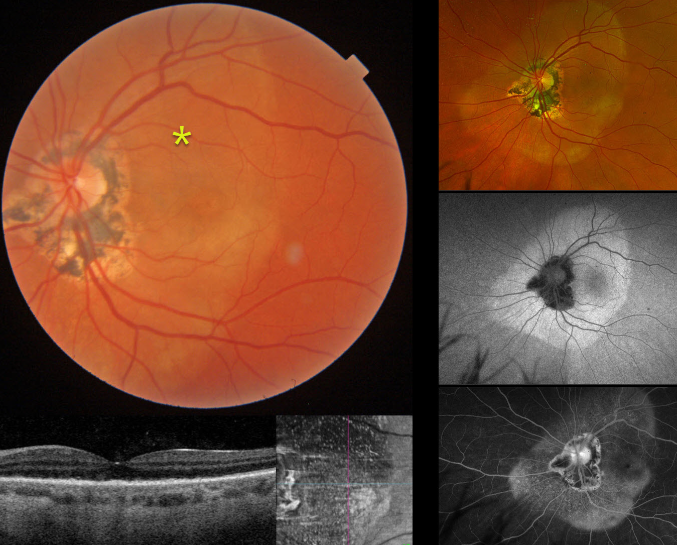
This 61YO female presented with 1 week of vision loss in her left eye. Vision was 20/25 in her normal OD and counting fingers OS.
Triton color imaging shows a subretinal yellow-white placoid lesion extending beyond the macula, along with ocular histoplasmosis-related peripapillary scarring. Swept-source OCT shows replacement of the outer retinal bands with hyperreflective material, which is seen as innumerable hyperreflective dots on en face imaging. Optos color RGB imaging shows the full extent of the placoid lesion. Fundus autofluorescence (FAF) of this lesion shows homogenous hyper-FAF. It diffusely stains on fluorescein angiography. Laboratory testing was positive for syphilis and negative for HIV. She was referred to an infectious disease specialist and placed on oral doxycycline 100mg BID due to a penicillin allergy. When last seen two and a half months after her initial presentation, and after completing a full 1-month course of antibiotics, vision improved to 20/40 with near-complete resolution of the retinal findings (not shown).
Learning Points:
Ocular manifestations of syphilis vary widely and include virtually all variations of anterior and posterior segment inflammation. Originally described by Gass (Ophthalmology 1990;97:1288-1297), placoid chorioretinitis is one of the more common presentations. Inflammation is localized to the outer retina and RPE and appears on OCT as disruption of the outer retinal bands and hyperreflective pyramidal lesions as seen in our patient (Hu et al, Ophthalmology Retina 2022;6:172-178). The outer retinal damage unmasks the underlying RPE, accounting for the hyperfluorescent placoid lesions.



