

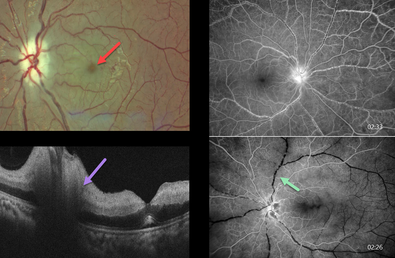
This previously healthy 14YO girl presented with 10 days of vision loss in her right eye. She also gave a history of recurrent migraine-type headaches. She saw an outside eye doctor, where blood pressure was not checked, and spectacles were prescribed. At presentation, vision was 20/100 OD and 20/20 OS.
Funduscopic examinations revealed mild bilateral disc edema with venous dilation (not shown). Blood pressure could not be detected in any limb. We suspected Takayasu arteritis and urgently referred her to a rheumatologist and neurologist. Unfortunately, she presented 2 weeks later with sudden vision loss in her left eye. Vision was 20/100 OD and no light perception OS.
Color photography shows a cherry red spot and pale disc edema in her left eye. Fluorescein angiography shows markedly delayed venous filling in both eyes at 2.5 minutes following injection. OCT scanning shows a swollen disc and marked inner retinal hyperreflectivity with posterior shadowing. An anterior chamber paracentesis was performed with no change in her funduscopic appearance.
Medical workup between our initial and subsequent examinations revealed no pulses in the radial and dorsalis pedis arteries, with reduced left femoral and left carotid pulses. Renal artery Doppler revealed high-resistance flow bilaterally in the main renal and segmental arteries with 80-90% lumen narrowing in the infrarenal aorta. Brain MRI revealed an old infarct with encephalomalacia and gliosis in the left cerebellar hemisphere. Immediately following her second exam with us, she was emergently admitted by rheumatology and given a methylprednisolone infusion. She was immediately lost to follow-up.
Learning Points:
Takayasu arteritis is an idiopathic granulomatous vasculitis involving medium- to large-sized vessels throughout the body. Involvement of the aortic arch and its branches can cause ocular ischemia, including anterior segment ischemia with rubeosis and neovascular glaucoma.
Posterior segment findings include retinal ischemia, neovascularization, and vascular occlusions. Surprisingly, over 50% of patients present with ocular findings as the initial manifestation of their disease. For a wonderful review of the posterior segment findings in Takayasu, see Sharma et al, Indian J Ophthalmol 2024;72:637-647.



