

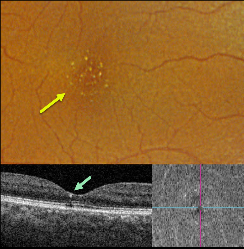
This 56YO female was referred for asymptomatic macular findings. The referring doctor mentioned that she may have retinal changes from the Tamoxifen that she was taking for over 3 years for breast cancer. Vision was 20/50 OD and 20/40 OS.
Optos color RGB imaging shows numerous tiny inner retinal crystalline deposits scattered throughout each fovea. These deposits appeared as tiny hyperreflective dots beneath the ILM on OCT B-scan and en face.
Our patient had already spoken with her oncologist before seeing us and had already stopped the Tamoxifen. The crystalline changes will likely remain and, hopefully, not progress going forward.
Learning Points:
Tamoxifen retinopathy shares phenotypes very similar to those of macular telangiectasia type 2 (MacTel 2, Lee et al., Ophthalmology Retina 2020;3:681-689), especially in the early stages, including retinal cavitations, right-angle venules, and inner retinal crystals.
The changes in Tamoxifen are confined to the central macula, whereas in MacTel2 they are present in a slightly larger area with an epicenter temporal to the foveal center (Hess et al, Ophthalmology Retina 2023;7:101-110). The retinal changes for both disorders likely share Muller cell dysfunction as the common cause.



