

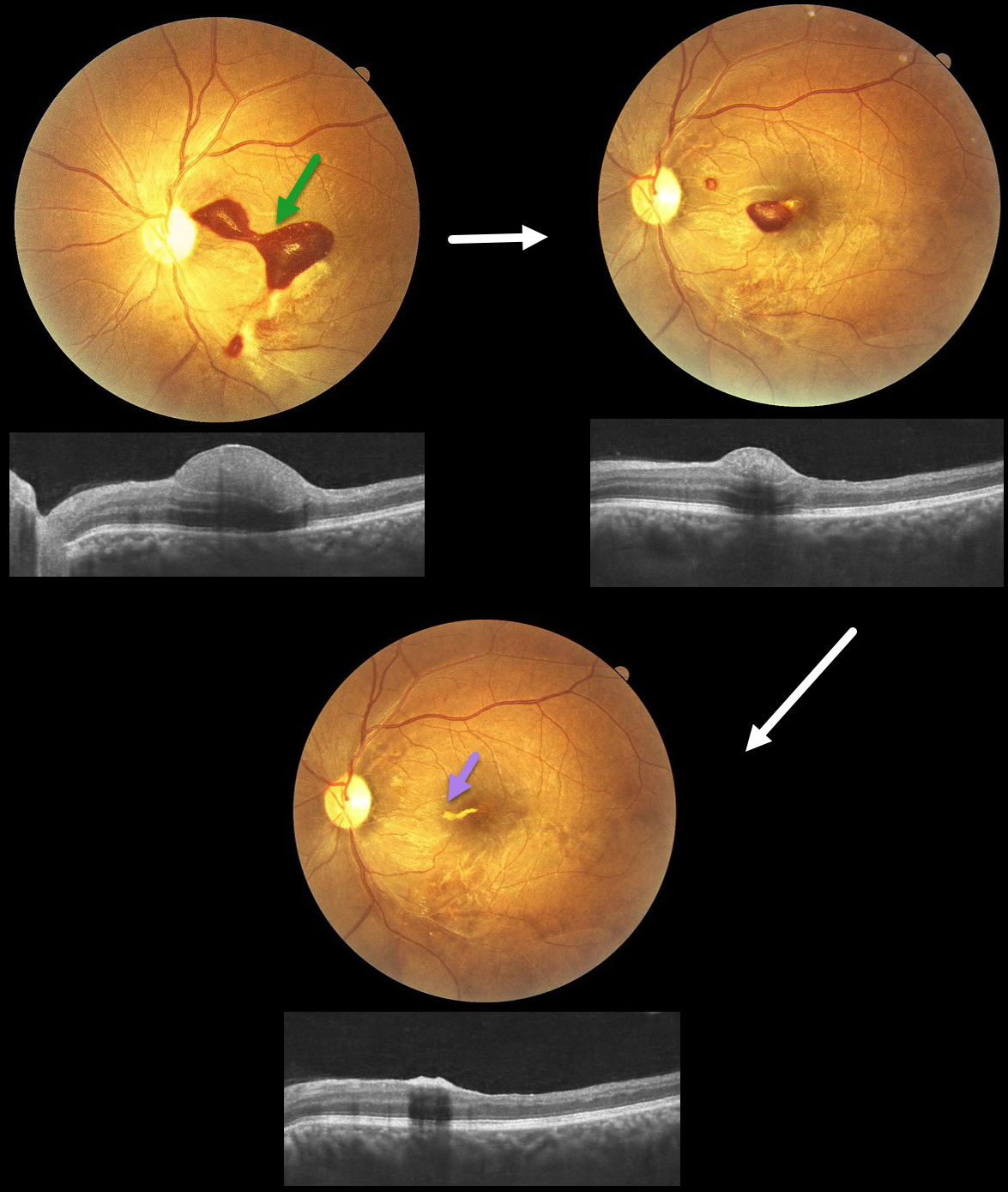
This 32YO male presented with 20 days of vision loss in his left eye. He underwent cardiovascular surgery 3 weeks earlier to remove a clot, followed 1 week later by a subarachnoid hemorrhage. Vision was 20/20 in his normal OD and 20/400 in his OS.
Fundus photography shows a subinternal limiting membrane (ILM) hemorrhage extending from the central macula, superonasally, towards the disc. This blood is hyperreflective on OCT. Observation was recommended.
One month later, vision improved to 20/70, with a marked decrease in sub-ILM blood. One month later, vision improved to 20/30, with near-complete resolution of the blood, except for a small amount of devitalized yellow blood extending nasally from the foveal center.
Learning Points:
Terson syndrome consists of intraocular hemorrhage (vitreous, subhyaloid, or retinal) associated with spontaneous or traumatic subarachnoid hemorrhage. Traditionally, the cause was attributed to elevated intracranial pressure (ICP), which caused cerebrospinal fluid effusion into the optic nerve sheath, compressing the central retinal vein and leading to subsequent rupture of retinal vessels. More recent evidence suggests glymphatic reflux, in which raised ICP causes subarachnoid blood to reflux through glymphatic channels into the globe (Kumaria et al, J Neurology 2022;269:1264-1271).



