

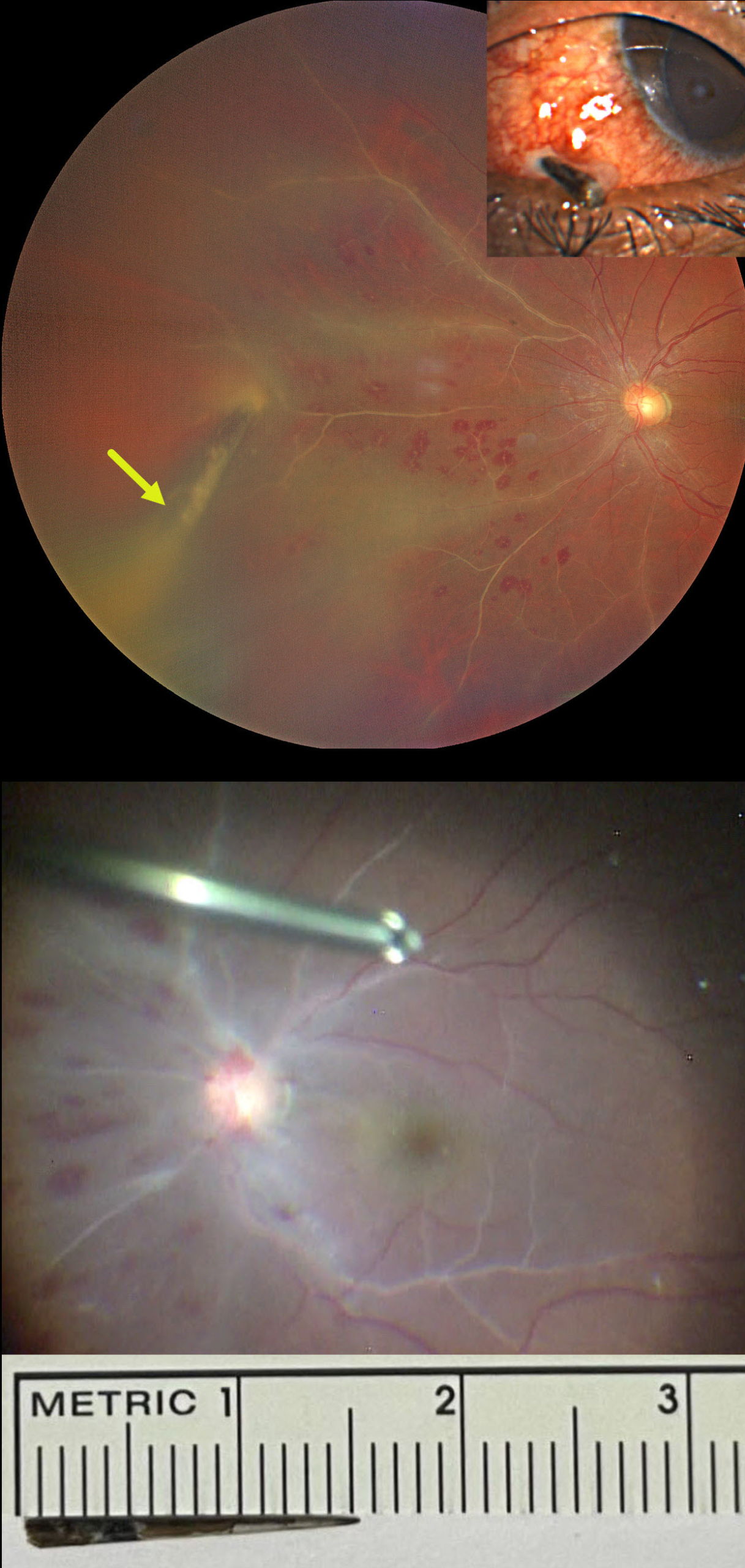
This 28YO male presented 5-6 hours following a perforating thorn injury to his left eye. He was standing in an open-moving loading vehicle when an Acacia tree branch suddenly hit his eye. Vision was 20/20 in his normal OD and 20/40 OS.
External examination showed a thorn lodged in the inferonasal sclera. There were moderate anterior chamber cells on slit lamp examination with a fibrinous pupillary membrane.
Fundus examination revealed vascular sheathing predominantly in the nasal quadrant, multiple, white-centered retinal hemorrhages, and vitreous exudates. The thorn penetrated the retina, extending into the vitreous cavity.
Despite being taken to surgery approximately two hours later, intraoperatively, there was a dramatic increase in vascular sheathing extending into the macula. Pars plana vitrectomy was performed with removal of the thorn foreign body, repair of the scleral laceration, cryotherapy, endolaser photocoagulation around the retinal break, silicone oil tamponade, and intravitreal antibiotic injection. The thorn measured approximately 15mm.
On the first postoperative day, the posterior segment appeared healthy with resolved vascular sheathing. One week postop, vision was 20/50 with a +5-diopter sphere, the retina remained completely attached, and good laser scarring was noted around the penetration site (not shown).



