

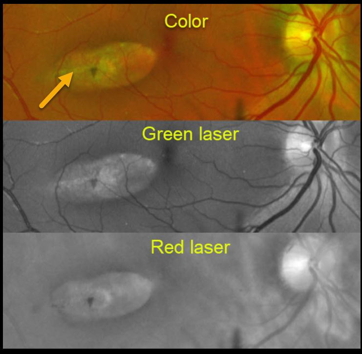
This patient has torpedo maculopathy.
This Optomap montage shows how the same lesion appears in color, green laser, and red laser.
OCT scanning, not available for this patient, would show attenuation of the RPE and outer retinal layers. Vision is almost always normal.
Learning Points:
Torpedo maculopathy was originally described by Gass as a solitary hypopigmented nevus of the RPE (Arch Ophthalmology 1992;1358-1359). These benign, unilateral lesions are teardrop-shaped and almost always located in the horizontal meridian of the temporal macula. There is a small risk of macular neovascularization.
These are congenital lesions with no systemic associations. For a great discussion to help differentiate torpedo maculopathy from other RPE lesions such as CHRPE and bear tracks, see Villegas et al, Ophthalmic Surg Lasers Imaging Retina 2014;45:222-226.



