

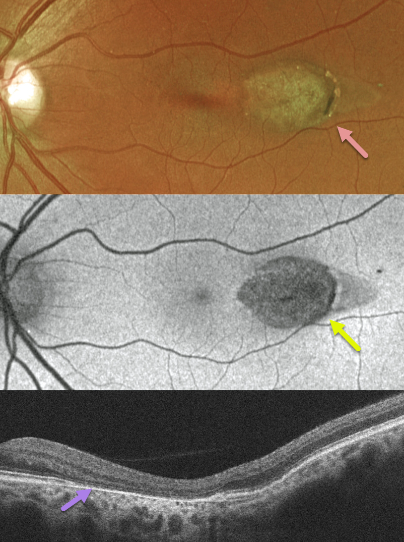
This healthy 44YO female was found to have this asymptomatic retinal finding in her left eye during a routine examination. The right eye was completely normal.
Optos color RG imaging shows a torpedo-shaped area of depigmentation in the temporal macula.
The lesion is hypo-autofluorescent. OCT scanning shows variable outer retinal and RPE atrophy.
Learning Points:
Torpedo maculopathy was originally described by Gass as a solitary hypopigmented RPE nevus (Arch Ophthalmology 1992;110:1358-1359). These benign, unilateral lesions are tear-drop shaped and are almost always located in the horizontal meridian in the temporal macula. Vision is almost always normal. There is a small risk of macular neovascularization.



