

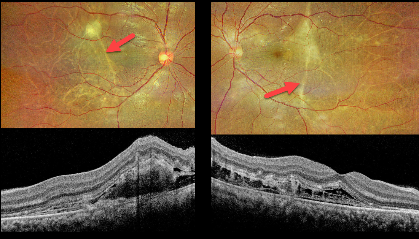
This 25YO female presented 10 days postpartum, complaining of bilateral blurred vision that began shortly following delivery, which was complicated by toxemia (preeclampsia). Blood pressure in our office was 180/90 mmHg. Vision was 20/30 bilaterally.
Optos color RG imaging shows multiple depigmented, wormlike subretinal tracks throughout each posterior pole. A larger contracting triangular-shaped lesion is noted in the superior left macula. Exudative fluid is present in the nasal maculas. OCT scanning shows outer retinal disorganization, an increased number of outer retinal bands, and pockets of variably hyperreflective fibrinous outer retinal fluid.
Learning Points:
Posterior segment findings in toxemia of pregnancy, which develop in up to one-third of patients, are primarily due to choroidal ischemia with secondary RPE dysfunction and exudative retinal detachments. Outer retinal ischemia can cause bizarre and pathognomonic OCT changes as seen in our patient (Song et al, Graefes Arch Clin Exp Ophthalmol 2013;251:2647-2650).
The clinical and OCT findings recover remarkably within weeks following normalization of blood pressure. Upon resolution, the fundi can appear normal or reveal telltale subretinal pigment clumps (Elschnig spots), streaks (Siegrist streaks), or broad areas of RPE pigmentary changes with intraretinal migration that can mimic prior blunt trauma or rhegmatogenous retinal detachment.



