

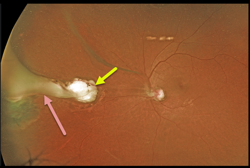
This 63YO female was referred for these asymptomatic unilateral fundoscopic changes. Optos imaging shows a midperipheral atrophic chorioretinal scar. Associated with this scar is a whitish fold of retina that extends anteriorly as a broader area of traction and scarring in the vitreous base.
A positive ELISA test can help to establish the diagnosis. Since our patient was asymptomatic, we did not pursue any further workup and recommended observation.
Learning Points:
Toxocariasis is a rare infection caused by the Toxocara canis roundworm, which matures in the dog intestine. The eggs are passed in stool, which can be ingested when young children come into contact with infected sand or dirt and then touch their mouths.
The ingested egg matures into a larva, which enters the systemic circulation from the intestine and can then migrate into the eye.
Death of the organism can cause variable findings, including endophthalmitis (typically a completely white and quiet eye), posterior granuloma, or peripheral granuloma.
Most commonly, in our experience, they present later in life as a triangular-shaped area of vitreous base organization/scarring, with a fold of retina extending more posteriorly.



