

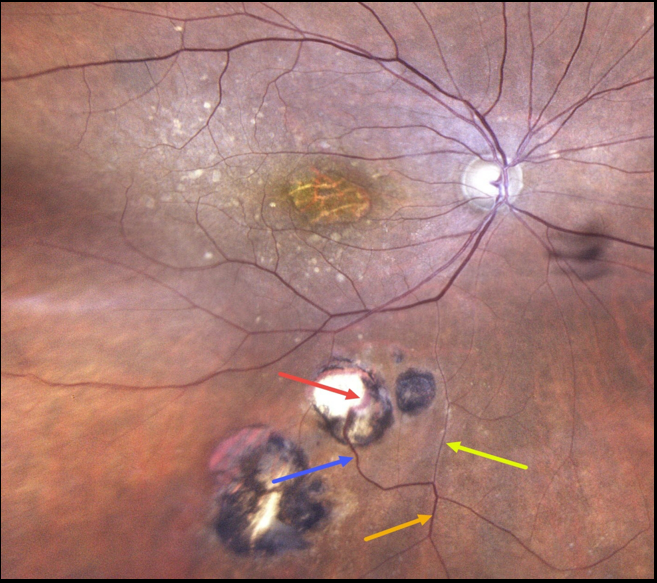
This 86YO female has a history of atrophic age-related macular degeneration with vision of 20/400 OD and 20/60 OS. Optos imaging of her right eye shows foveal atrophy with surrounding mixed drusen.
Multifocal inactive toxoplasmosis scars are noted inferiorly. An inferior retinal vein bifurcates, with a markedly attenuated branch continuing on its path towards the optic nerve. The other branch has a more normal caliber, dives into one of the toxoplasmosis scars, and drains into the choroid.
Learning Points:
Under normal conditions, the retinal and choroidal circulations remain separate and distinct. Rarely, the two circulations connect directly through a chorioretinal anastomosis.
These can occur in chorioretinal scars (typically from inactive toxoplasmosis lesions, as in this patient), in macular telangiectasia, and in disciform scars.



