

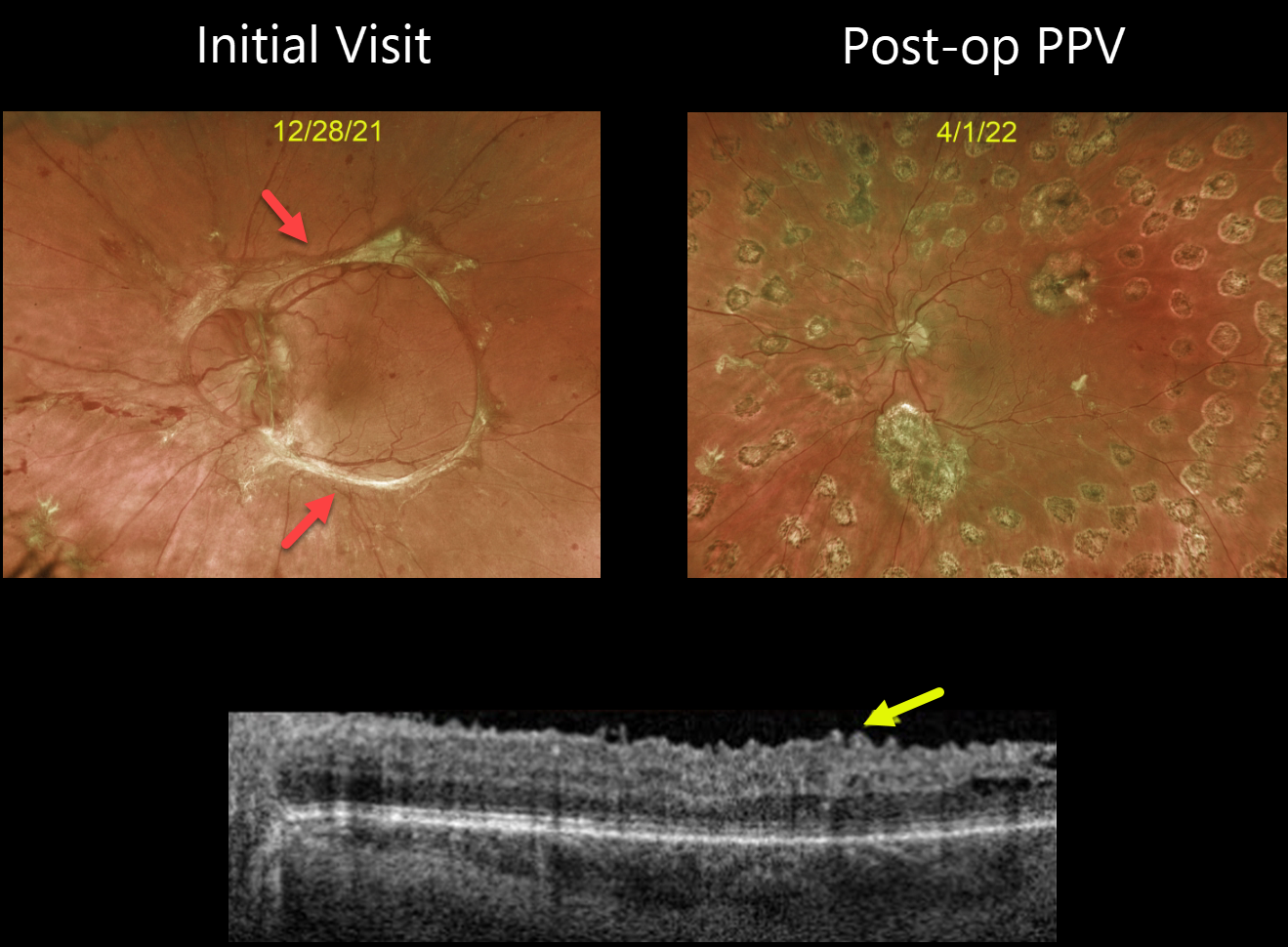
This 32YO female with type 1 diabetes presented with an 18-month history of vision loss in her left eye. Vision was counting fingers.
Optos imaging beautifully captures the pre-operative photos of the tractional retinal detachment, caused primarily by fibrosed retinal neovascularization extending in a circular pattern from the nerve along the arcades.
The second image shows the patient four months following vitrectomy. Swept-source OCT shows an irregular inner retinal contour, which is common following these vitrectomies.
Vision improved to 20/50. She is currently undergoing panretinal photocoagulation in her contralateral eye.



