

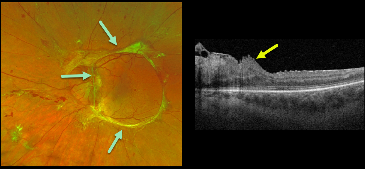
This 32YO female with type 1 diabetes presented with an 18-month history of vision loss in her left eye. Vision was counting fingers.
Optos imaging beautifully captures the traction retinal detachment from mostly fibrosed retinal neovascularization extending circularly from the nerve along the arcades.
Triton swept-source OCT surprisingly shows relatively mild macular distortion despite the severe vision loss, which is most likely due to vitreopapillary traction.
Intravitreal Avastin followed by vitrectomy will be performed.
Learning Points:
Tractional retinal detachment is one of the main complications of untreated or incompletely treated proliferative diabetic retinopathy.



