

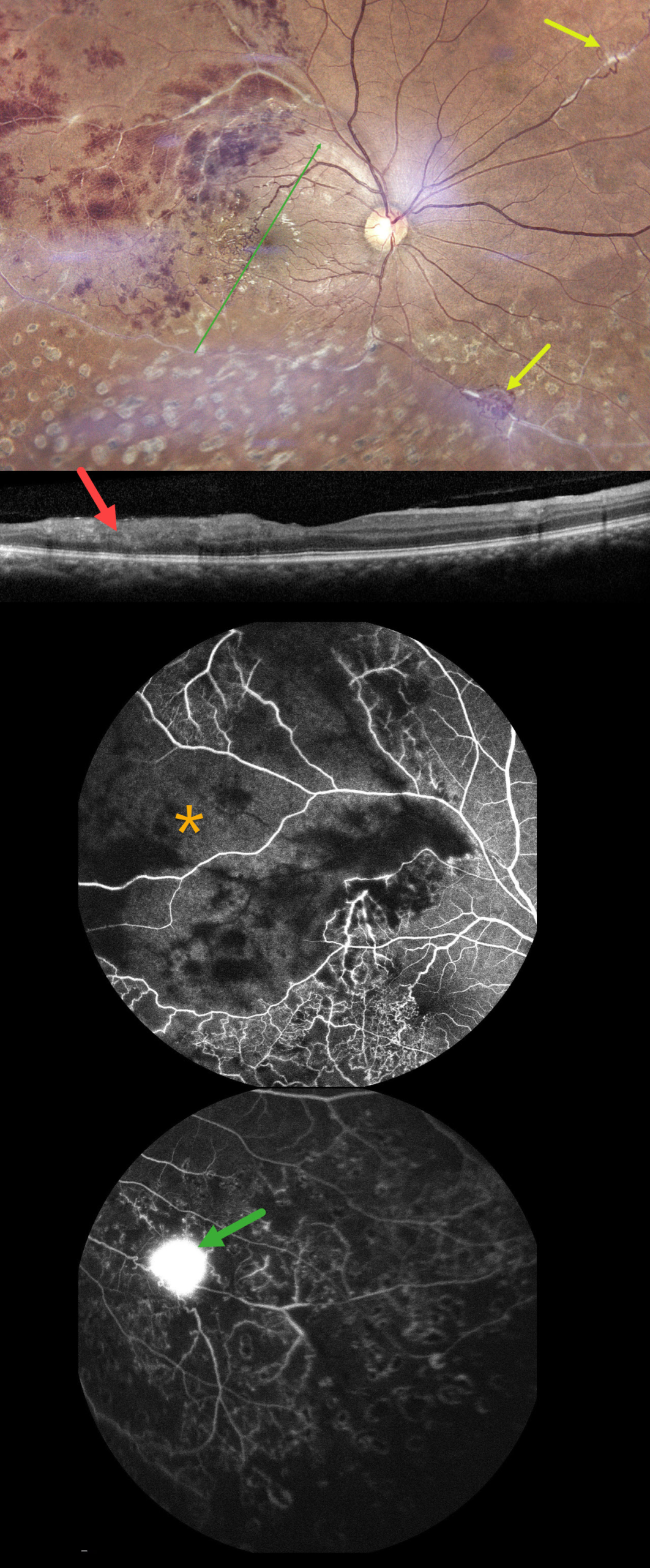
This 40YO male presented with 10 days of vision loss in his right eye. There was a history of prior laser treatment to this eye elsewhere. Vision was 20/40 OD and 20/20 in his normal OS.
Fundus photography shows superotemporal retinal hemorrhages consistent with a branch retinal vein occlusion (BRVO), with a possible additional BRVO inferotemporally. Collateral vessels are noted in the temporal fovea with scatter laser scarring in the inferior hemiretina. Skip vasculitis can be seen in the superotemporal quadrant along with venous sheathing in other areas as well. There are a few areas of fibrosed peripheral neovascularization (NV). OCT scanning shows variable temporal macular thinning with disorganization of retinal inner layers (DRIL). Fluorescein angiography shows profound ischemia within the superotemporal BRVO, along with vascular leakage and peripheral ischemia. Leakage from the Inferonasal NVE can be seen in the late stages.
Systemic workup was consistent with Ocular Tuberculosis (TB), including a positive Mantoux skin test (12mm), positive Quantiferon TB testing, and calcified lung nodules on contrast-enhanced computed tomography. He was started on oral steroids and antitubercular therapy, and scatter laser was planned for the areas of angiographic ischemia.
Learning Points:
The differential for occlusive peripheral retinal vasculitis (Huvard et al, Ophthalmology Retina 2022;6:43-48) includes TB, rheumatologic disorders/systemic vasculitides (granulomatosis with polyangiitis, Bechet’s disease, systemic lupus, etc.), idiopathic retinal vasculitis and neuroretinitis, and sarcoidosis. Historically, Eales disease has been used to describe a variety of heterogeneous disorders, including tuberculous vasculitis, and, due to its ambiguity, we prefer not to use this term.



