

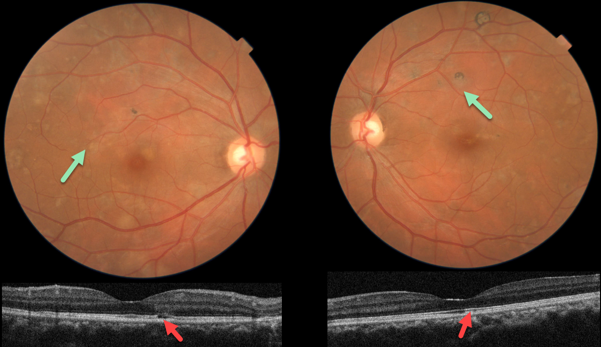
This 38YO male presented with floaters in each eye. He denied any past medical history. Vision was 20/20 OU.
Fundus photos show multifocal patches of variably pigmented chorioretinal scarring throughout each posterior pole.
OCT scanning in each eye shows variable outer retinal atrophy and ellipsoid disruption. Unfortunately, fundus autofluorescence and fluorescein angiography are not available.
Subsequent workup was consistent for active tuberculosis, including a positive QuantiFERON-TB Gold and chest x-ray findings. Systemic anti-tuberculous therapy was begun.
Learning Points:
Serpiginous choroiditis is an idiopathic, usually bilateral chorioretinal inflammatory condition most commonly found in middle-aged men (Khanamiri and Rao, Surv Ophthalmology 2013;58:203-232).
Recurrent areas of inflammation develop along the edge of a previous scar, producing scarring that meanders from the optic nerve outwards. Vision is good unless scarring or macular neovascularization extends through the macular center.
Tubercular serpiginous-like choroiditis mimics serpiginous, but affects younger patients with more multifocal and peripheral recurrences and progression if untreated (Agarwal et al, AJO 2020;220:160-169).
Although the exact mechanism remains unclear, a direct or indirect infectious trigger by Mycobacterium tuberculosis is believed to cause choroiditis.



