

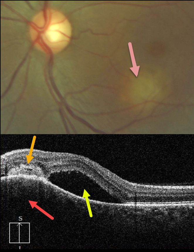
This 33YO female presented with this symptomatic lesion in her left macula. Vision was 20/25 in her normal right eye and 20/300 in her left eye.
A granulomatous lesion is noted in the inferior left macula. A vertical OCT B-scan through this lesion shows choroidal hyporeflective thickening, an overlying subretinal/intraretinal hyperreflective lesion, and superior subretinal fluid.
Laboratory testing was positive for QuantiFERON-TB Gold, and negative for FTA, CBC, and ACE. Unfortunately, despite numerous attempts to reach this patient, she was lost to follow-up.
Learning Points:
Tuberculosis (TB) is a systemic disease caused by Mycobacterium tuberculosis, characterized by the formation of caseating granulomas throughout the body. Although pulmonary involvement is the most common, extrapulmonary sites can involve the gastrointestinal, skin, cardiovascular, genitourinary, and central nervous systems, as well as the eyes.
Ocular involvement, similar to syphilis, can mimic virtually any type of uveitis, including anterior, intermediate, posterior, and panuveitis, retinitis and retinal vasculitis, neuroretinitis, optic neuropathy, and choroidal granuloma, choroiditis, and scleritis.
For an excellent review of intraocular tuberculosis, see Bupta et al, Survey Ophthalmology 2007;52:561-587.



