

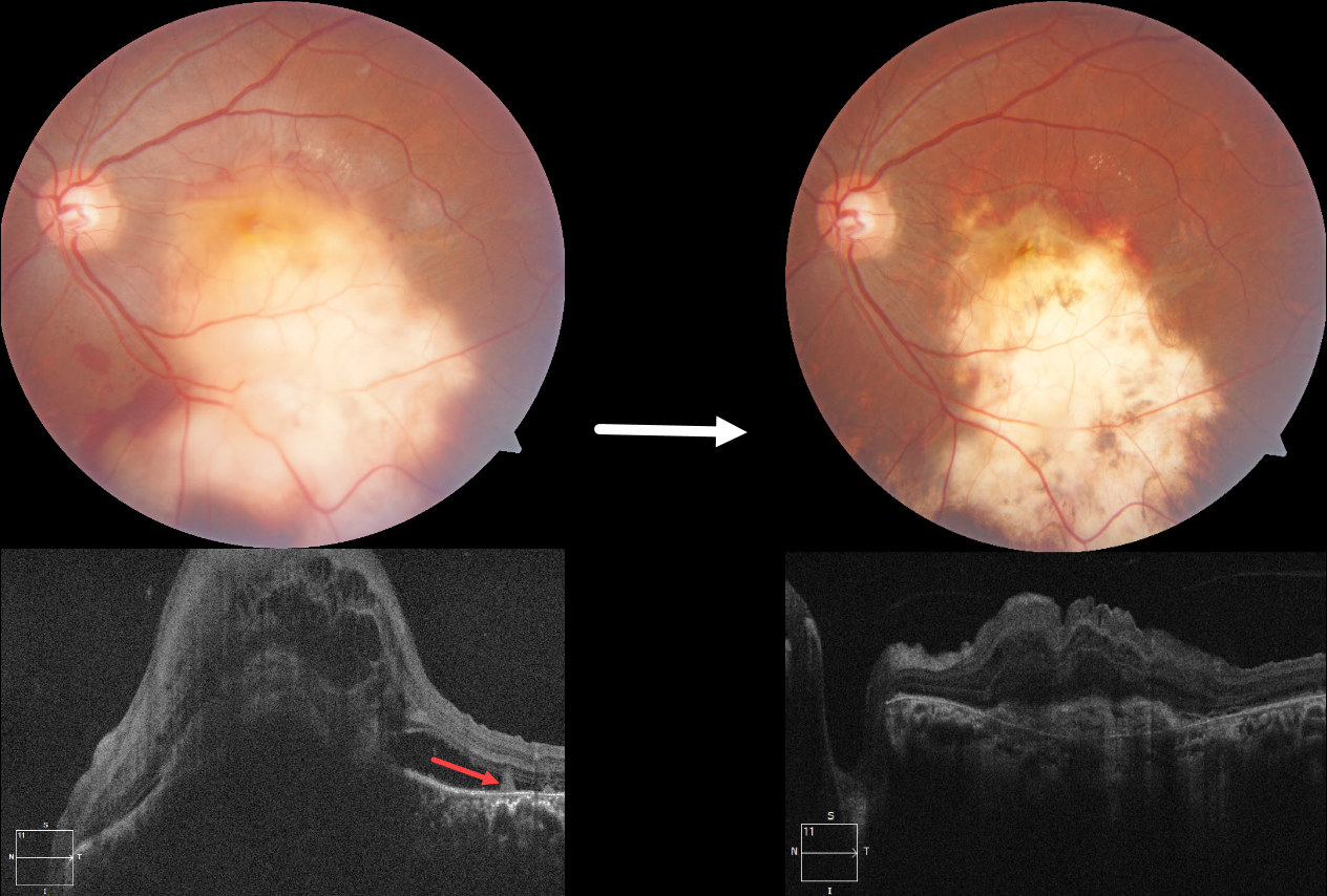
This 36YO male presented with 10 days of vision loss in his left eye. One month earlier, he was diagnosed with disseminated tuberculosis (TB), peritonitis, and pleural effusion, and was placed on systemic antitubercular therapy (ATT). Vision was 20/20 in his normal right eye and counting fingers in his left eye.
Fundus photography shows a large elevated yellowish submacular lesion that extends into the inferior midperiphery. Subretinal blood is noted along its superior and inferior margins. Faint lipid is present superotemporally, and some retinal striae are seen temporally. Spectral domain OCT shows a large dome-shaped hyporeflective choroidal lesion with overlying cystic retinal thickening, subretinal fluid, and subretinal hyperreflective material. A few hyperreflective pyramidal lesions are noted above the RPE temporally.
We started him on oral corticosteroids. In view of the vascularized lesion with subretinal blood, an intravitreal injection of Avastin was also given. Six weeks later, there is a relatively flat scar with near complete resolution of the blood. The choroidal lesion has resolved with an overlying hyperreflective subretinal scar and decreased retinal thickening. Unfortunately, vision remained at counting fingers.
Learning Points:
Although TB most commonly presents with pulmonary involvement, extrapulmonary sites can include the gastrointestinal, skin, cardiovascular, genitourinary, and central nervous systems, including the eyes. Ocular involvement, like syphilis, can mimic virtually any type of uveitis, including anterior, intermediate, posterior, and panuveitis, retinitis and retinal vasculitis, neuroretinitis, optic neuropathy, choroidal granuloma, choroiditis, and scleritis.
For an excellent review of intraocular tuberculosis, see Bupta et al, Survey Ophthalmology 2007;52:561-587.
Tubercular granulomas can be distinguished from sarcoid lesions by their solitary nature, yellow color, larger size, and vascularization (Agarwal et al., AJO 2021;226:42-55). All these features were found in our patient. Vascularization is thought to arise from localized hypoxia, leading to increased levels of vascular endothelial growth factor. Anti-VEGF injections and systemic corticosteroids, in addition to ATT, are helpful in hastening granuloma resolution and reducing secondary exudation (Lee et al, Graefe’s 2022;260:1641-1650).



