

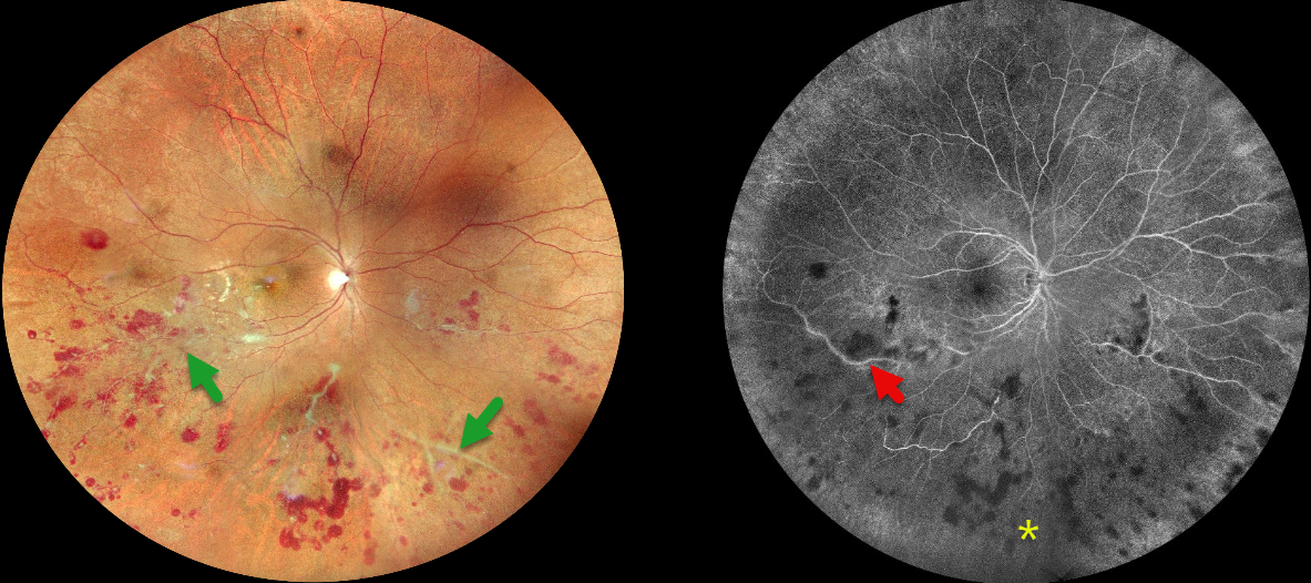
This 27YO male presented with 10 days of floaters and photophobia in his right eye. He was tentatively diagnosed with tuberculosis (TB) several days earlier and sought a second opinion with us. Recent Mantoux skin testing was markedly positive (20 mm induration), and QuantiFERON-TB Gold was positive as well. Vision was 20/20 bilaterally.
Fundus photography shows inferior and nasal vascular sheathing, inner and deep retinal hemorrhages in the inferior hemisphere, and focal areas of whitish inner retinal ischemia in the inferior macula. Fluorescein angiography shows variable midperipheral ischemia with focal vascular staining. The left eye was normal.
Chest CT was supportive of active pulmonary TB, and the patient was started on antitubercular treatment (ATT) by the pulmonary team on the day of our exam.
When he returned four days later, his vision was 20/40 OD. We recommended an intravitreal anti-VEGF injection followed by scatter laser, but unfortunately, he was then lost to follow-up.
Learning Points:
The differential for occlusive peripheral retinal vasculitis (Huvard et al, Ophthalmology Retina 2022;6:43-48) includes TB, rheumatologic disorders / systemic vasculitides (granulomatosis with polyangiitis, Bechet’s disease, systemic lupus, etc), idiopathic retinal vasculitis and neuroretinitis, and sarcoidosis. Historically, Eales’ disease has been used for a variety of heterogeneous disorders, including tuberculous vasculitis, and due to its ambiguity, we prefer not to use this term.
The diagnosis of tubercular vasculitis is often presumptive and difficult to diagnose without isolation of TB from ocular tissue or fluids. However, as with our patient, it is a reasonable diagnosis in the proper clinical setting, including in those from endemic areas and with a systemic diagnosis of TB (Agrawal et al, 2017 37:112-117). Systemic ATT, along with local ocular treatment for neovascular complications (anti-VEGF injections, scatter laser photocoagulation), is recommended.



