

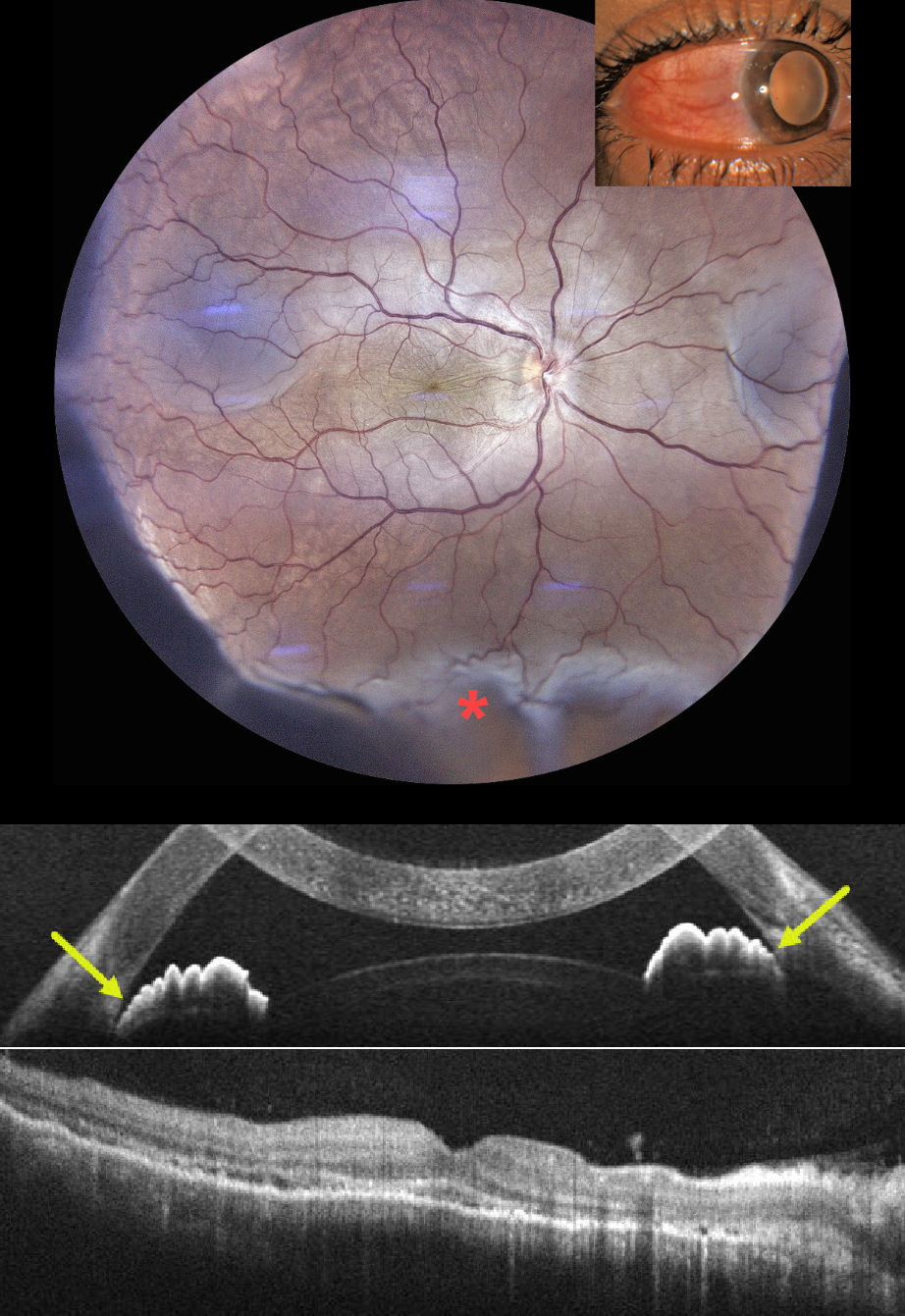
This previously healthy 35YO male presented with 1 week of pain, redness, and decreased vision in his right eye. He had been treated by an outside doctor for conjunctivitis. Vision 20/200 OD and 20/20 in his normal OS. Slit lamp examination showed temporal scleral injection. Intraocular pressure was 23 mm Hg.
Color photography shows radiating chorioretinal folds throughout the macula with an inferior exudative retinal detachment. OCT scanning of the macula shows chorioretinal folds, with anterior chamber OCT showing a narrow-to-closed angle. B-scan ultrasonography showed subtenon’s fluid (not shown).
Following a 3-week course of tapering oral steroids and glaucoma drops, vision improved to 20/40 with reduced chorioretinal folds and complete resolution of the exudative detachment. Rheumatologic workup was negative, but Mantoux skin testing and QuantiFERON-TB GOLD were positive, with a subsequent diagnosis of latent tuberculosis (TB). Antituberculous therapy was recommended, but the patient refused treatment.
Learning Points:
Tuberculous posterior scleritis is a rare manifestation of ocular TB (Agarwal and Majumder, Indian J Ophthalmol 2019;67:1362-1365), which itself occurs in approximately 1.5% of patients with confirmed systemic TB. Ocular findings include optic disc edema, choroiditis, and sclerochoroidal thickening with widening of the sub-Tenon space. It may occur via direct hematogenous dissemination of Mycobacterium tuberculosis or through a hypersensitivity-mediated immune response to the organism and can develop even in the absence of clinically active pulmonary disease.
Diagnosis, as in our patient, is largely presumptive, requiring a high index of suspicion supported by bloodwork, chest imaging to assess pulmonary involvement, and exclusion of mimics such as sarcoidosis, syphilis, and metastatic disease.



