

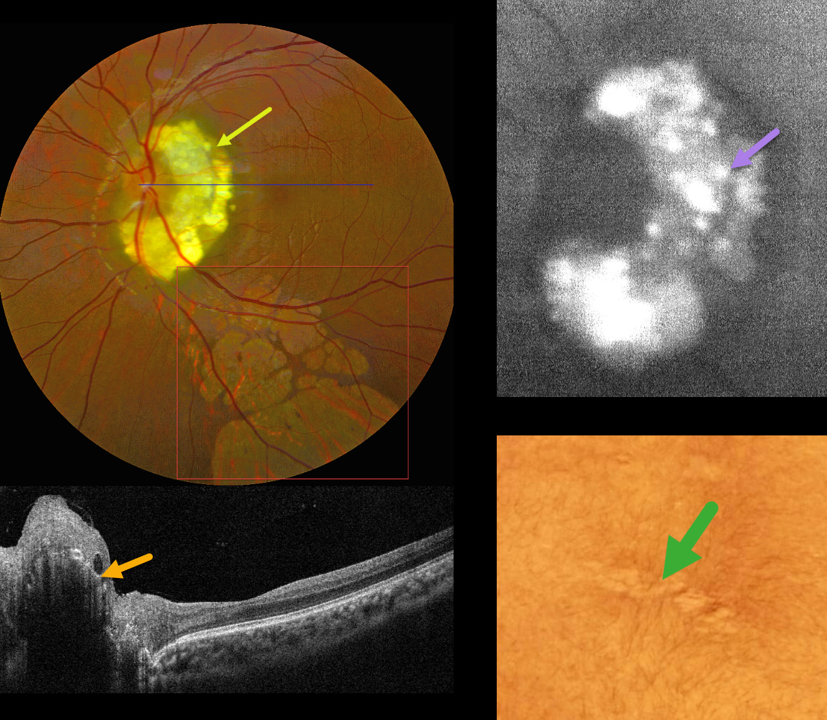
This 17YO male presented with a known history of tuberous sclerosis, childhood seizures, and decreased vision in his left eye. Vision was 20/20 in his normal OD and 20/40 OS.
Color photography shows a glistening yellow-white inner peripapillary retinal lesion surrounding the superior, temporal, and inferior edge of the optic nerve, composed of innumerable semi-transparent spherules which hyper-autofluoresce. OCT scanning shows an elevated, hyperreflective inner retinal lesion with a hyporeflective intralesional cavity. An area of skin depigmentation (ash leaf spot) was noted on his lower back. There are numerous variably sized and variably confluent lobules of subretinal depigmentation extending from the macula inferiorly.
Learning Points:
The best-known retinal finding in tuberous sclerosis is retinal astrocytic hamartoma. Early in life, these inner retinal lesions have a fleshy white appearance and are often multifocal and bilateral. With time, as in this patient, they show a more classic “mulberry appearance” with significant calcification. The calcified lesions, as in our patient, hyper-autofluoresce while the flat, semitransparent lesions found earlier in life are hypoautofluorescent. Rarely, the vascular component can cause intraretinal exudation requiring treatment.
Retinal achromic patches, although much less described in the literature, are found in up to 39% of tuberous sclerosis patients (Rowley et al, Br J Ophthalmology 2001;85:420-423). They are thought to be analogous to the hypopigmented skin lesions and can appear in one or both eyes, singly or in multiples, and are typically located in the retinal periphery. Unliked astrocytomas, these lesions are flat and subretinal.



