

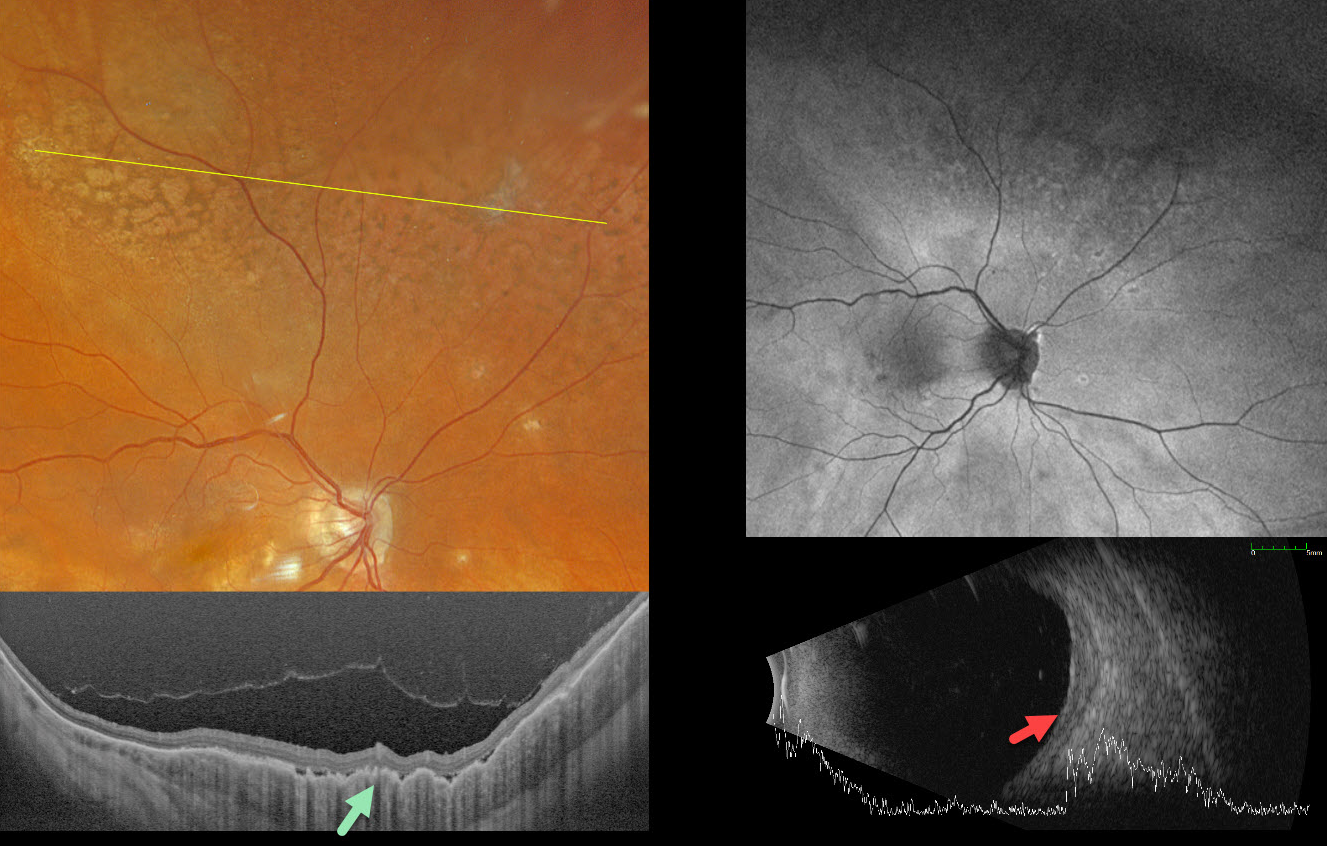
This 69YO female was receiving ongoing bilateral anti-VEGF therapy for neovascular age-related macular degeneration when she presented with new asymptomatic fundus changes in her left eye. There was no significant past medical history.
Optos color RGB imaging shows a leopard spot pattern of superior midperipheral pigmentary changes. Silverstone swept-source OCT shows a markedly thickened choroid with overlying chorioretinal folds. Fundus autofluorescence (FAF) is fairly unremarkable despite these funduscopic findings. The choroid is diffusely thickened on B-scan ultrasonography, with a maximal thickness of 1.90mm.
CT of the chest, abdomen, and pelvis was negative. MRI of the brain and orbits was also negative. We felt this represented primary uveal lymphoma, and this was confirmed with an outside third opinion before beginning serial intravitreal methotrexate injections. After 11 months of treatment, the choroid flattened clinically and on ultrasonography, shrinking to 1.09mm in thickness (not shown).



