

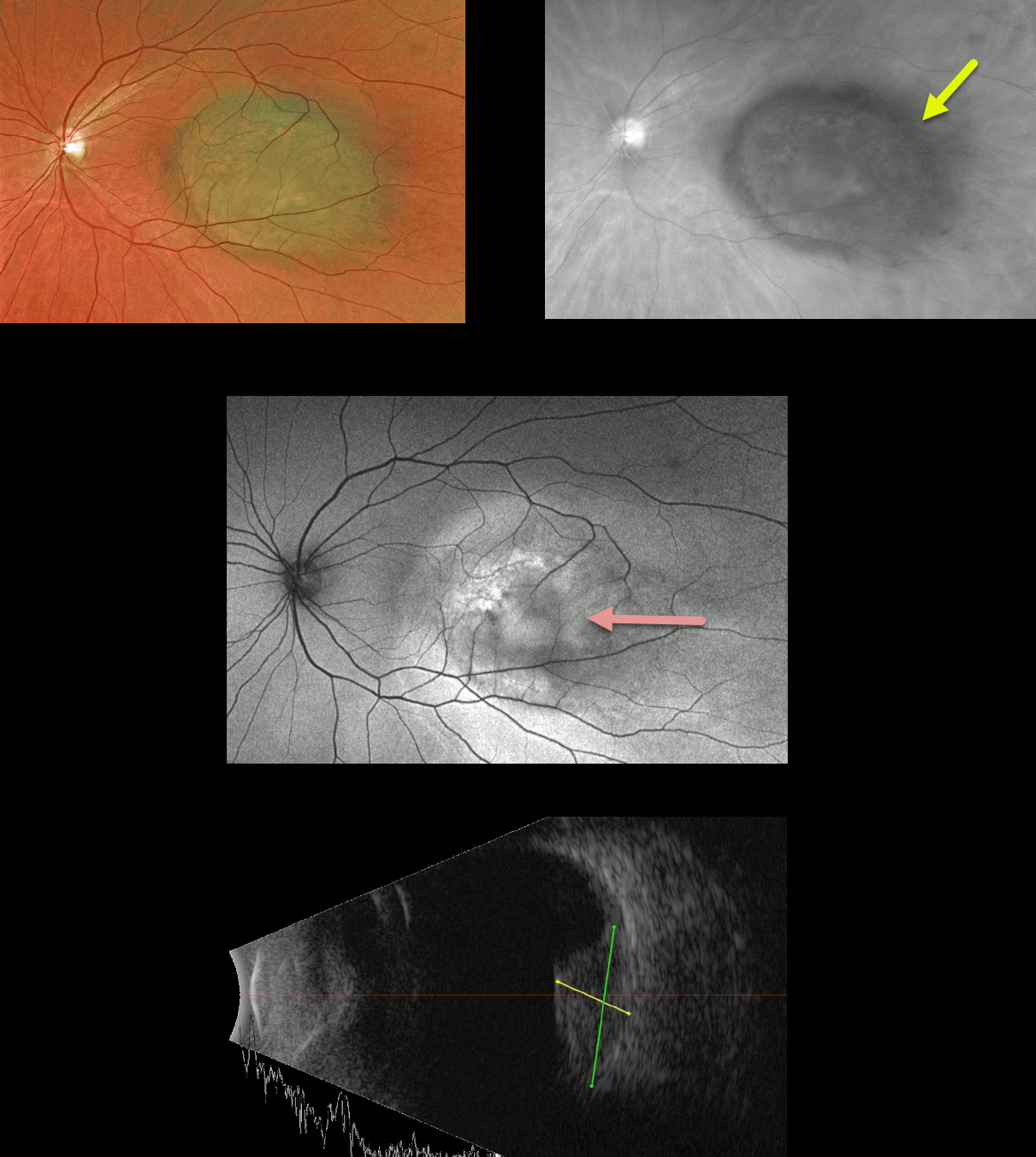
This 52YO female presented with 20/200 vision in her left eye due to a uveal malignant melanoma. Amazingly, she was completely asymptomatic since she was right eye dominant.
The hyperpigmented lesion is well visualized on the Optos red channel and exhibits variable hyper- and hypoautofluorescence. The tumor measured about 5.5mm in thickness and 12mm in basal diameter on B-scan.
Radioactive plaque therapy was recommended.
Learning Points:
Clinical characteristics of choroidal nevi at risk for progressing to a uveal MM were described by Shields et al. (Ophthalmology 1995;102:1351-1361). The most recent version of this famous acronym, To Find Small Ocular Melanoma Doing Imaging (TFSOM-DIM, Retina 2019;39:1840-1851) is:
To (Thickness >2mm)
Find (Subretinal Fluid)
Small (Symptoms, vision <= 20/50)
Ocular (Orange pigment)
Melanoma (Melanoma hollow)
DIM (DIIaMeter >5mm)



