

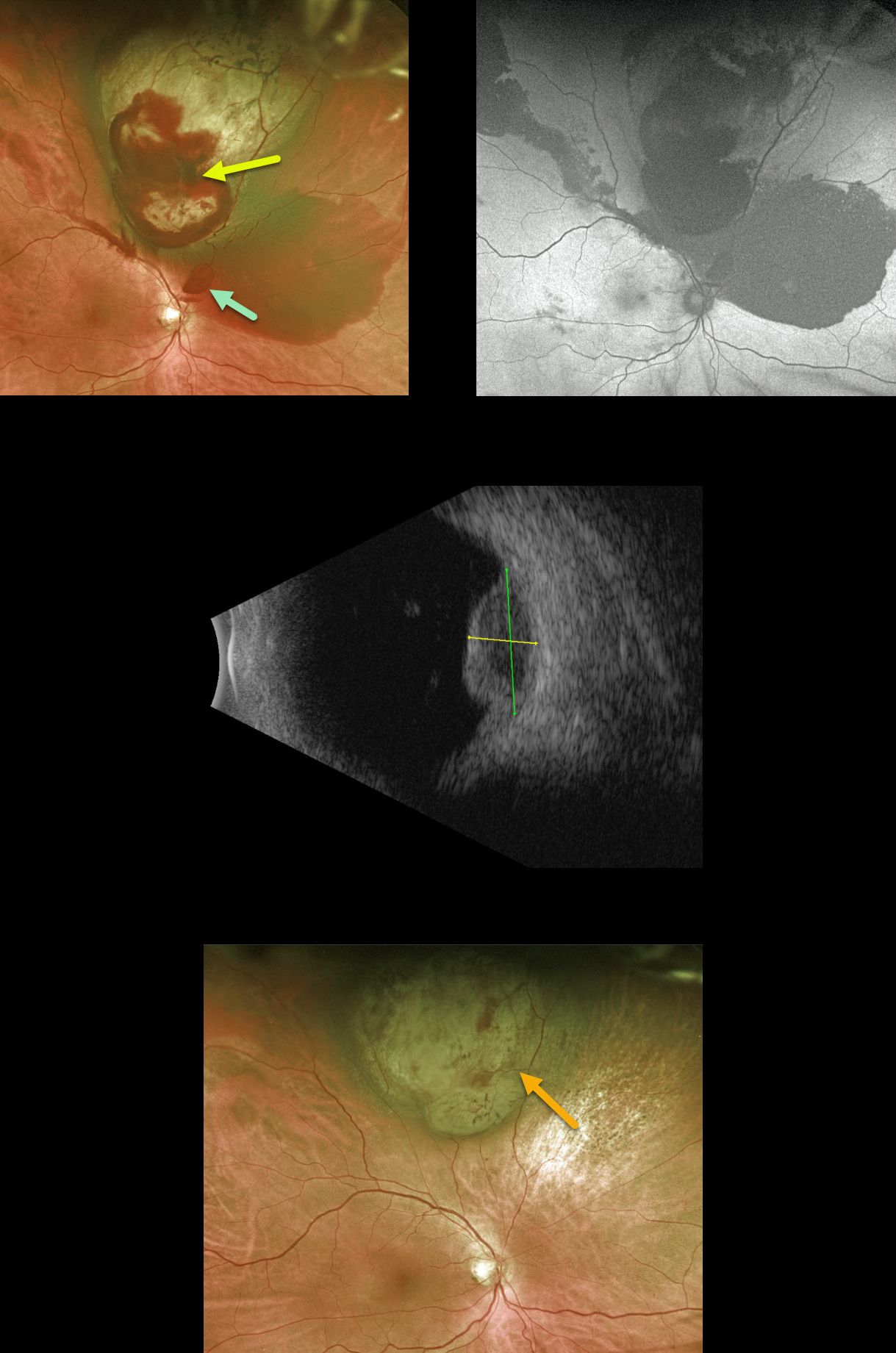
This 60YO male presented with 20/25 vision in his right eye despite a superior uveal malignant melanoma. The tumor was associated with subretinal blood overlying and outside the lesion, as well as a small area of preretinal blood just superior to the optic nerve.
Fundus autofluorescence showed no lipofuscin. The tumor measured 5.55mm in thickness, with a basal diameter of 11.07mm, and appeared hollow on ultrasound, while the overlying subretinal blood was more echogenic.
A few days prior to radioactive plaque therapy, he noted a severe worsening of vision and was found to have a dense vitreous hemorrhage, obscuring the view of the tumor (not pictured).
Pars plana vitrectomy was performed at the time of plaque placement to clear the view and allow for transvitreal fine-needle aspiration.
About 3 months postoperatively, the tumor regressed to a thickness of 3.5mm. The subretinal blood resolved with secondary pigmentary changes, and his vision returned to baseline.
Learning Points:
Genetic expression profiling demonstrated a Class 1B, PRAME-negative tumor.
The patient continues to be followed closely for local recurrence and metastatic disease.



