

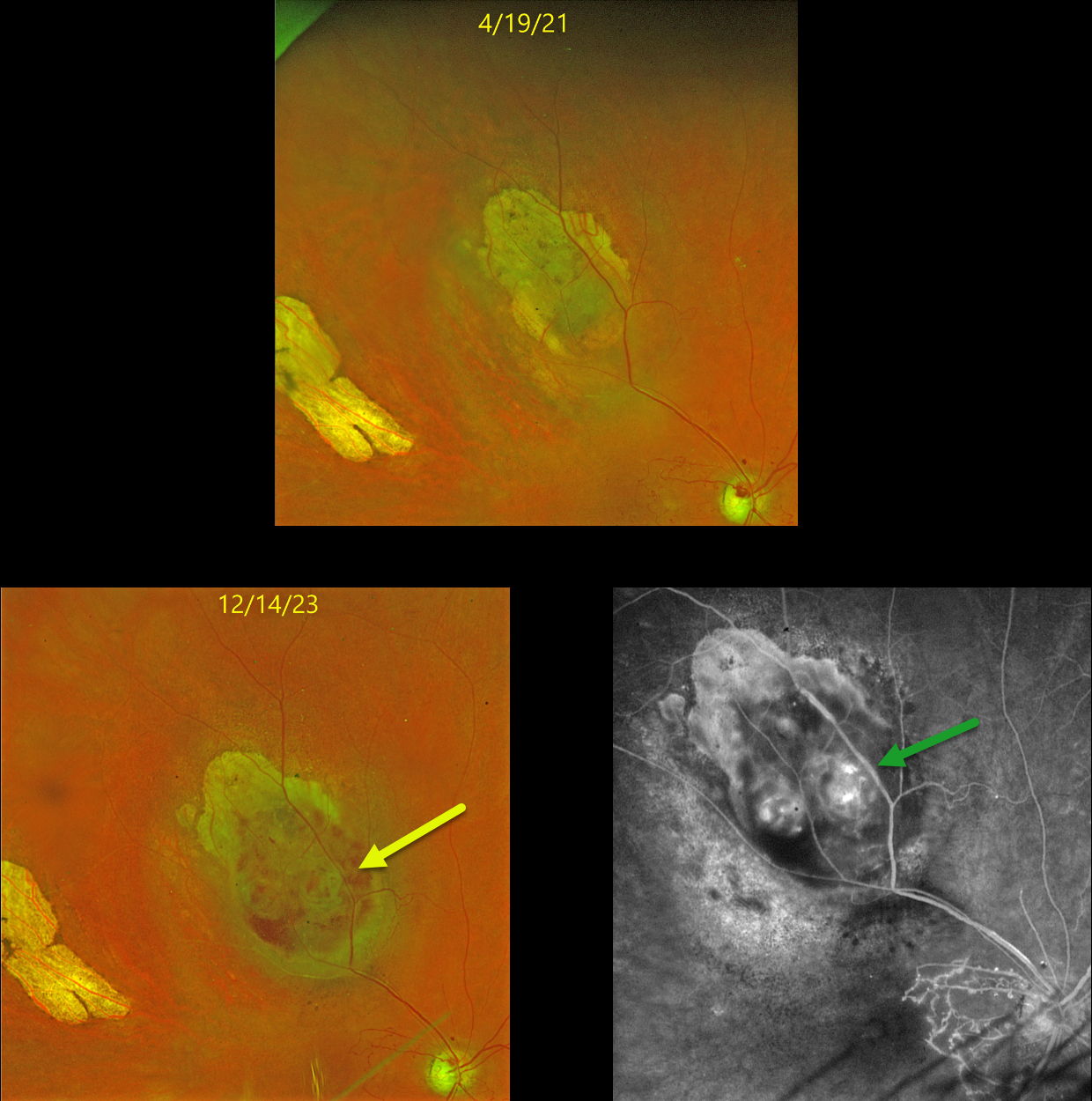
This 59YO female underwent iodine-125 plaque therapy in 2010 for a uveal malignant melanoma (MM) in her right eye. The tumor initially regressed and has remained stable with yearly examinations since. Vision has been stable at counting fingers due to a secondary branch retinal vein occlusion and radiation retinopathy.
In June 2023, she presented with new floaters and vision loss in this eye from a new moderate vitreous hemorrhage coming from the MM. Vision was light perception. Once the blood cleared following an intravitreal Avastin injection, the tumor showed some hemorrhages within or anterior to the lesion on Optos color RGB imaging.
Fluorescein angiography showed intrinsic vascularity and profound macular capillary loss. B-scan ultrasonography revealed a nodular appearance with heterogeneous echogenicity (not shown). There was definite growth compared with the tumor size several years earlier. Metastatic workup was negative. After extensive discussion, the patient elected for enucleation, which was scheduled in the near future.
Learning Points:
The Collaborative Ocular Melanoma Study (COMS) reported a 10% 5-year local treatment failure rate following plaque radiation (Jampol et al., Ophthalmology 2002;109:2197-2206). Our patient’s failure developed about 13 years following brachytherapy and reminds us of the need for long-term surveillance of these treated tumors.



