

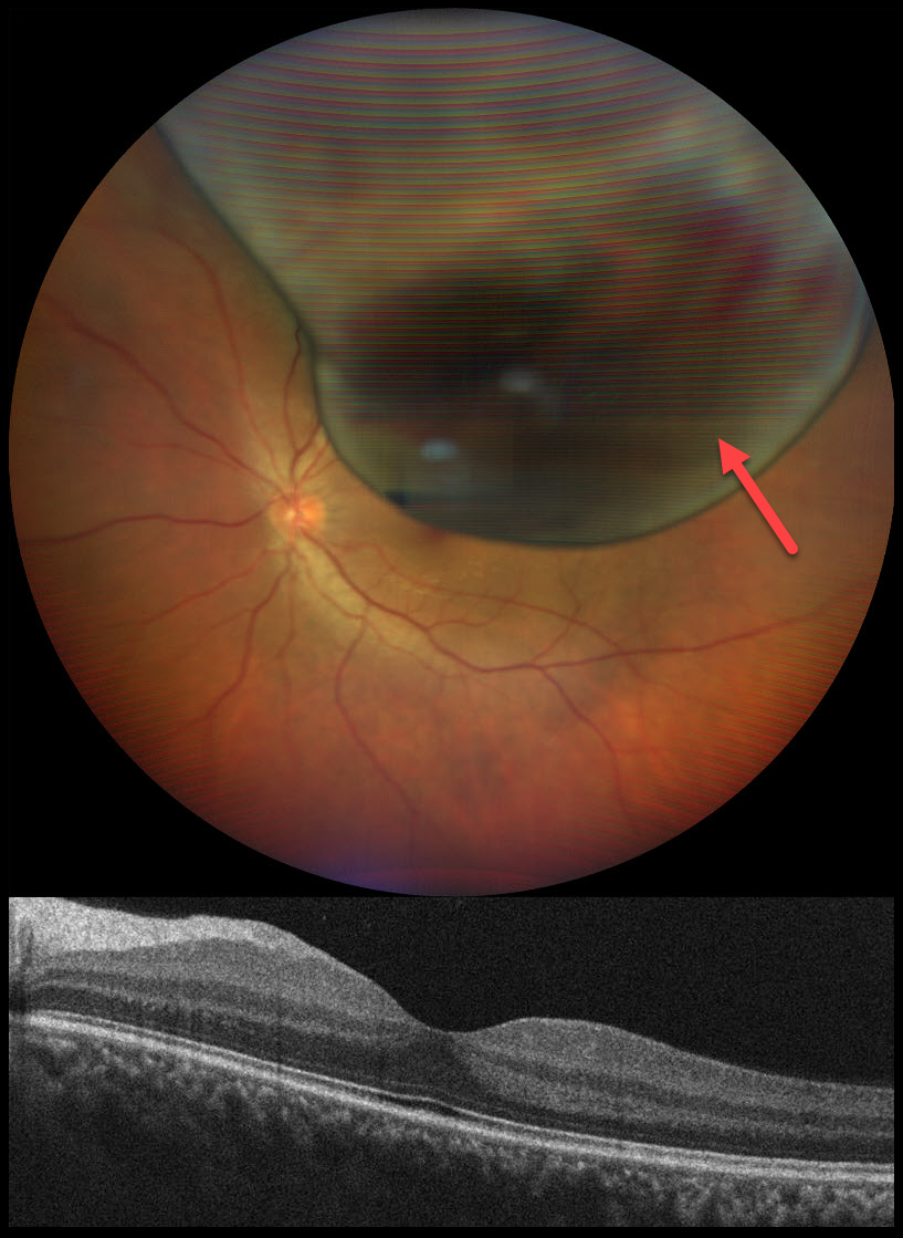
This 32YO male presented with a peripheral shadow of vision in his left eye for several weeks. Vision was 20/20 OU.
Color photography shows a large, elevated, well- defined, pigmented choroidal mass with a few overlying hemorrhages. Despite the lesion nearly draping over the central macula, foveal OCT scanning is remarkably normal. He was immediately referred to an ocular oncologist for a presumed uveal malignant melanoma. He was immediately lost to follow up.
Learning Points:
Uveal melanoma is a malignant tumor arising from melanocytes in the uveal tract (iris, ciliary body, or choroid). The choroid is the most common site, accounting for 90% of uveal melanomas. Uveal melanoma is the most common primary intraocular cancer in adults, whereas metastatic disease is the most common intraocular malignancy. The liver is the most common metastatic site, although metastatic disease is rare on presentation. Metastases are more common with large tumors, BAP1 mutations, and certain tumor-related genetic markers (including positive PRAME and Class 2).
Despite our patient having normal vision, their left eye harbored a blinding and potentially lethal uveal MM. This reminds us why we must always listen to our patient and explain their symptoms even with a normal acuity.



